in vitro modeling of skin photoaging: Development of evaluation tools for cosmetics
IFSCC 2014, 27th congress, Paris
BOUDIER D.(1), SOLINGEAS N.(1), MARCHAND L.(1), BORDES S.(1), CLOSS B.(1), BARRAULT C.(2), PEDRETTI N.(2), GARNIER J.(2), BERNARD F.X.(2)
(1) SILAB, ZAC de la Nau, 19240 Saint-Viance, France,
(2) Bioalternatives, 1 bis rue des Plantes, 86160 Gençay, France,
INTRODUCTION
Dermatologists and cosmetic scientists are always interested in standardized biological models for better understanding the skin modifications at the tissue, cellular and molecular levels under different conditions (age, chemical, physical, microbiological stresses…). Repetitive UV exposure of the skin is well known to elicit photo-aging mainly consisting of wrinkling and sagging the skin. [1]
AIM: Develop in vitro models of growing complexity and analysis of the relevance of these in vitro evaluation tools to better understand the cutaneous photo-aging process, particularly at the dermis level.
MATERIAL & METHODS
MODEL 1: COMPARISON BETWEEN NORMAL HUMAN DERMAL FIBROBLASTS (NHDF) AND NHDF AFTER REPETITIVE UVA IRRADIATIONS (NHDFuv)
- NHDF were cultured for 4 days and maintained in culture for additional 24h or 96h.
- NHDF were cultured and irradiated with 15 J/cm² UVA once a day, for 4 days, obtaining NHDFuv, and maintained in culture for additional 24h or 96h.
- Expression of COL1A1, MMP-1 and MMP-3 by RT-qPCR
- Procollagen I and MMP-1 release by ELISA
MODEL 2: COMPARISON BETWEEN A RECONSTRUCTED SKIN (RecSkin) WITH NHDF (RecSkinF) AND A RECSKIN WITH NHDFUV (RecSkinFuv)
- NHDF were introduced in the dermal compartment during the reconstruction of the 3D skin model: RecSkinF.
- NHDFuv were introduced in the dermal compartment during the reconstruction of the 3D skin model: RecSkinFuv.
- Histological analysis after Hematoxilin/Eosin coloration
- Expression of COL1A1, MMP-1 and MMP-3 by RT-qPCR
- These analyses were performed at D6 and D13 of the reconstruction
MODEL 3: COMPARISON BETWEEN A RECSKIN (RecSkinCtrl) AND A RECSKIN SUBMITTED TO DIFFERENT DOSES AND TYPES OF REPETITIVE UV IRRADIATIONS (RecSkinUV)
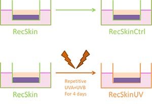
- RecSkin built with NHDF was used as a control: RecSkinCtrl. (Figure 1)
- RecSkin built with NHDF was submitted to UVA+B irradiations at different doses, once a day, for 4 days: RecSkinUV. (Figure 1)
- Histological analysis after Hematoxilin/Eosin coloration
- Transcriptomic analysis by Affimetrix chip
- Expression of COL1A1, COL3A1, MMP-1, MMP-3, MKI67, CDKN1A and CDKN2A by RT-qPCR
- Procollagen I, MMP-1 and MMP-3 release by ELISA
- Immunohistological staining of procollagen I and visualization by fluorescence microscopy
Figure 1. Protocol
RESULTS
MODEL 1: COMPARISON BETWEEN NORMAL HUMAN DERMAL FIBROBLASTS (NHDF) AND NHDF AFTER REPETITIVE UVA IRRADIATIONS (NHDFUV)
A strong repression of COL1A1 transcript (>3-fold) in NHDF versus NHDFuv was measured. The aged phenotype was confirmed by the strong over-expression of MMP-1 (>5-fold) and MMP-3 (>12-fold) 24h post-irradiation. Interestingly, the same trend was also observed at the protein level since the release in the culture media of procollagen I was decreased (>2-fold) and of MMP-1 was increased (>2-fold).
MODEL 2: COMPARISON BETWEEN A RECONSTRUCTED SKIN (RecSkin) WITH NHDF (RecSkinF) AND A RECSKIN WITH NHDFUV (RecSkinFuv)
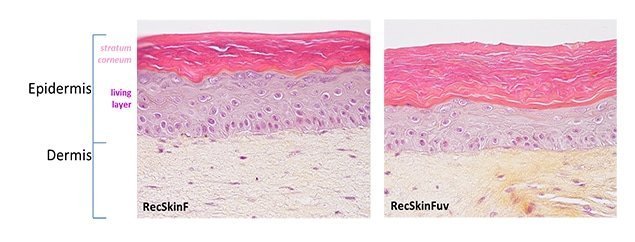
Figure 2. Histological analysis of RecSkinF in comparison with RecSkinFuv
Compared to RecSkinF, the histological analysis of RecSkinFuv epidermis showed a slight loss of cohesion, less thickness of the living layers and less organized basal layer. (Figure 2) Modulations of COL1A1, MMP-1 and MMP-3 expression in RecSkinFuv comparatively to RecSkinF were less pronounced than those observed in monolayer cultures before their introduction in the RecSkin (data not shown).
MODEL 3: COMPARISON BETWEEN A RECSKIN (RecSkinCtrl) AND A RECSKIN SUBMITTED TO DIFFERENT DOSES AND TYPES OF REPETITIVE UV IRRADIATIONS (RecSkinUV)
3.1. HISTOLOGICAL ANALYSIS
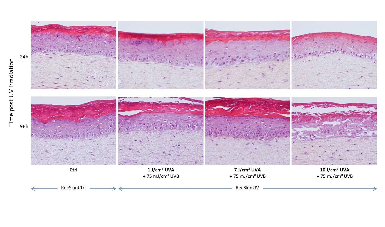
Figure 3. Histological analysis of RecSkinCtrl in comparison with RecSkinUV
3.2. TRANSCRIPTOMIC ANALYSIS
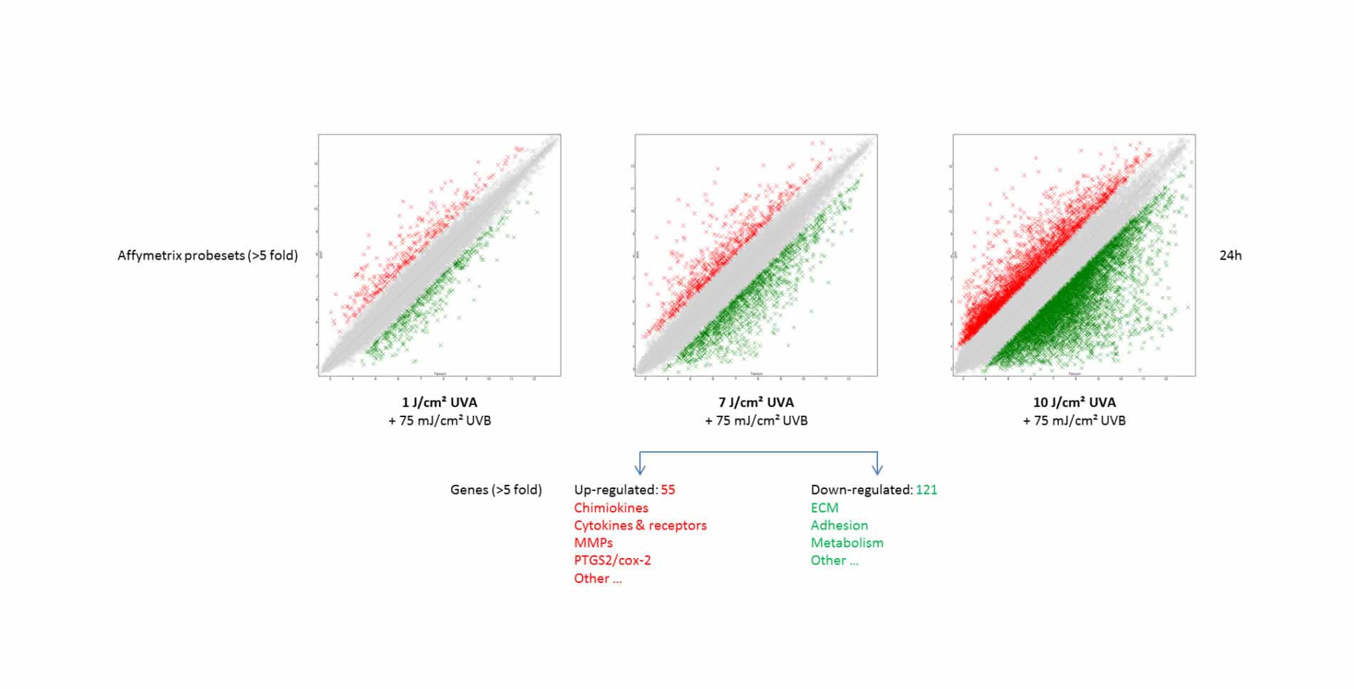
Figure 4. Transcriptomic analysis of RecSkinUV at different doses of repetitive irradiations in comparison with RecSkinCtrl
Histological analysis of ResSkinUV in comparison to RecSkinCtrl showed that a UVB background (75 mJ/cm²) associated with the lowest dose of UVA (1 J/cm²) had very few effect on the tissue morphology, 24h and 96h post- irradiations (corresponding respectively to the D9 and D12 of the reconstruction). Interestingly, the increasing doses of UVA from 7 J/cm² to 10 J/cm² led to morphological modifications of the tissue aspect, in particular at the epidermal level with the appearance of spongiosis. The lesions were dramatic with UVA at the maximum dose of 10 J/cm², particularly 96h after the latest irradiation. (Figure 3)
The comparison of gene expression profile between RecSkinCtrl and RecSkinUV 24h post-irradiations showed an overall modulation (up and down) of key skin biomarkers. (Figure 4) By focusing on the intermediary condition of repetitive UVB (75 mJ/cm²) + UVA (7 J/cm²) exposure, we highlighted an up-regulation of 55 genes involved in number of essential functions (inflammation, matrix degradation…) and a down-regulation of 121 genes important in extracellular matrix construction, cell adhesion, metabolism… The condition of repetitive irradiations at the doses: UVB (75 mJ/cm²) + UVA (7 J/cm²) was retained for further investigations.
3.3. QUANTITATIVE ANALYSIS OF GENE EXPRESSION AND PROTEINS SYNTHESIS
- Aging biomarkers
The comparative analysis by RT-qPCR between RecSkinCtrl and RecSkinUV (UVB 75 mJ/cm² + UVA 7 J/cm²) 24h post-irradiations showed that MKI67 (marker of proliferation KI67) expression was down-regulated. We underlined also that gene expression of CDKN1A (cyclin-dependent kinase inhibitor 1A) and CDKN2A (cyclin-dependent kinase inhibitor 2A) was up-regulated in RecskinUV. These two genes code for proteins named p21 and p16, which are known in the literature as biomarkers of aging. [2]
- Extracellular matrix biomarkers
Figure 5. Quantitative analysis of gene expression of extracellular matrix biomarkers in RecSkinUV (UVB 75 mJ/cm² + UVA 7 J/cm²) 24h and 96h post-irradiations and in comparison with RecSkinCtrl
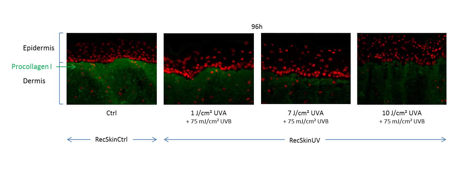
Figure 6. Study of procollagen I synthesis by fluorescence immunohistology in RecSkinUV compared to RecSkinCtrl
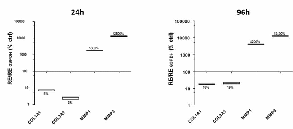
Quantitative measurements of modulations observed previously by transcriptomic analysis were carried out by RT-qPCR, fluorescence immunohistology and ELISA methods. Key matrix molecules like collagen I and III were strongly down-regulated in RecSkinUV, at the transcriptional level. (Figure 5) The same trend was visualized at the protein level by fluorescence immunohistology of procollagen I. (Figure 6) Then, the procollagen I release in the culture medium of RecSkinUV was also strongly diminished (measurement by ELISA method, data not shown) in comparison to RecSkinCtrl. Inversely, genes coding for biomarkers involved in the degradation of extracellular matrix like MMP-1 and MMP-3 were up-regulated in RecSkinUV in comparison to RecskinCtrl, at the transcriptional level (Figure 5) and these results were confirmed at the protein level by ELISA assessment in the culture media (data not shown).
CONCLUSION
For the design of the most reliable in vitro model to study early biological events occurring after repetitive UV exposure and responsible for the appearance of skin photo-aging clinical signs, our experiment plan consisted in testing:
– different in vitro cutaneous biological models, from classical monolayer cell cultures to 3D models composed of the two main skin compartments: the epidermis and the dermis;
– different conditions of repetitive UV exposure that had not to induce irreversible damages but susceptible to provoke an accelerated in vitro photo-aging process;
– different key biological markers, at both transcriptional and protein levels, known in the literature for their major role in the skin metabolism and the aging process. [3]
Under the different conditions of this study, we demonstrated that the most relevant in vitro model was the reconstructed skin (RecSkin) submitted to repetitive UV irradiations once a day, for 4 days with UVB (75 mJ/cm²) + UVA (7 J/cm²) during the reconstruction (RecSkinUV).
Only under these conditions, we showed modulations of both aging markers and key biomarkers of skin metabolism, without altering drastically the 3D model.
This novel approach gives a new scientific insight about the photo-aging process and proves that the 3D “compromised” skin models are interesting as in vitro evaluation tools for the design of effective molecules to prevent or to treat the skin photo-aging.
References:
[1] Mathes S.H., Ruffner H. and Graf-Hausner U., The use of skin models in drug development., Adv. Drug Deliv. Rev., (2014) 69-70, 81-102.
[2] Chen Q .M., Replicative Senescence and Oxidant-Induced Premature Senescence: Beyond the Control of Cell Cycle Checkpoints. Annals of the New York Academy of Sciences, (2000) 908, 111-125.
[3] Gilc hrest B.A., Photoaging., J. Invest. Dermatol., (2013) 133,(E1): E2-6.
Related posts
Check out Bioalternatives’ updates and experience new testing ideas
- Bioassays, models and services
- Posts and publications
- Events


