


Transmission electron miscroscopy
Transmission electron microscopy (TEM) enables the high resolution (1 to 2 Å) observation of very thin biological samples containing small compounds, such as cellular organelles etc. This microscopy technique also makes it possible to analyse the composition of the sample at the atomic scale.
A beam of electrons produced by an electron gun is transmitted through the sample at high speed. The interaction of the electrons and the sample generates a radiation, which results in an image that can be viewed from a computer monitor. This optical image is in black and white and shows different contrasts according to the quantity of the electrons that have travelled through the sample.
Examples of images obtained by TEM
Scanning electron microscopy
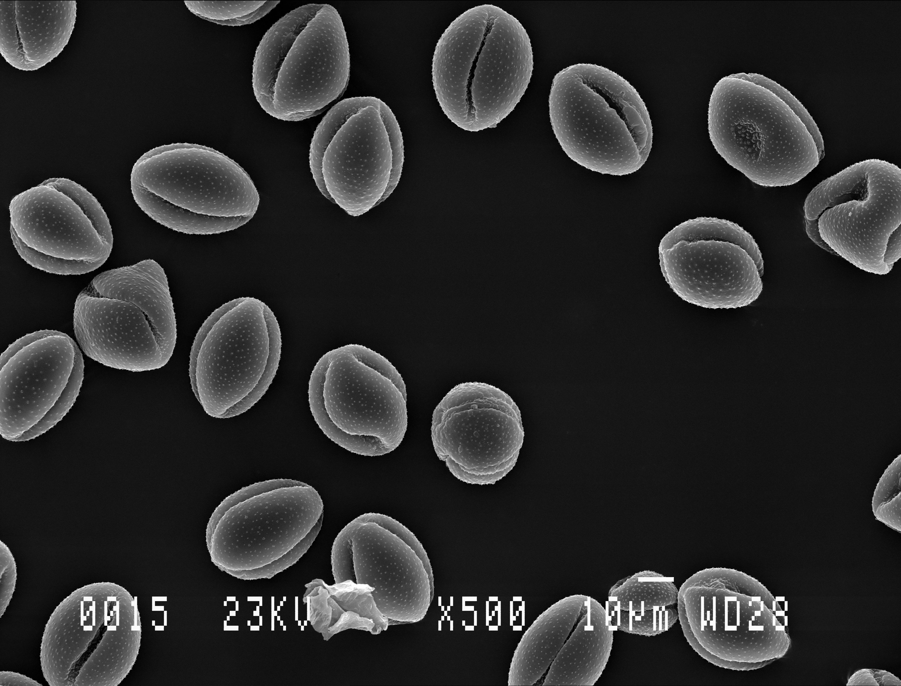
Scanning electron microscopy (SEM) enables the observation of large-scale biological samples at the nanometer scale.
In contrast to TEM, the electron beam does not travel through the sample but scans across its surface. The reflected radiations produce a signal and an image, viewable from a computer monitor, is thus generated. This image is in black and white and shows different contrasts, which provide a 3D vision of the surface with a high level of detail (e.g. visualization of bacteria on the skin or characterization of the hair surface condition).
Examples of images obtained by SEM


