Assays to Study the Hallmarks of Skin Aging
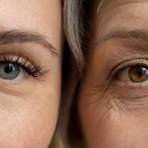
The skin is a window into the aging process: easy to observe, it reflects the cumulative effects of time, genes, the environment, and lifestyle. To describe this natural aging process, we can talk about chronological age (years), biological age (the ability of skin cells to function and repair themselves), and perceived age (the appearance we project).
Throughout our lives, external aggressions cause tissular and cellular damage, including DNA damage, oxidative stress, and the breakdown of collagen and elastin, among the most prominent. All of this results in well-known clinical signs such as wrinkles, loss of elasticity, thinning, but also in chronic inflammation or metabolic disorders that can slow down skin repair and promote further aging signs.
The biological processes that underpin skin aging and longevity are closely linked to the 12 hallmarks of aging, identified by López-Otín et al. (2023). These hallmarks help us connect visible signs of aging to biological processes occurring at the cellular and molecular levels, and thus contribute to identifying biomarkers, testing interventions and treatments, and advancing research in skin longevity.
At QIMA Life Sciences, we offer customized tools and services to investigate and address each of these hallmarks.
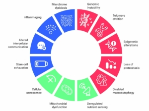
Explore Our Skin Aging Solutions for the Primary Antagonistic Integrative Hallmarks Below
Click on a category to explore our solutions
Genomic Instability:
The exposure to UV rays, pollution or oxidative stress constantly expose the genome of the cells to a variety of stress factors. These can lead to mutations in the DNA which compromise the genetic integrity of the cells. To maintain their functionality, cells usually rely on internal repair mechanisms. As we age, these systems can become less effective, allowing genetic damage to accumulate. This buildup participates in the normal aging process but also increases vulnerability to age-related diseases.
OUR SOLUTIONS
- Quantify CPDs – UV-induced DNA lesions
- Detect pATM (phospho-ATM) – DNA double‑strand break response signaling
- Measure 8-OHdG – Oxidative DNA damage
- Detect HP1α – Heterochromatin marker
- Detect γH2AX – DNA double‑strand break marker
- Analyze mtDNA copy number, deletions & heteroplasmy – Mitochondrial genome integrity
- Assess histone carbonylation – oxidative modifications affecting chromatin
- Perform the Comet Assay – DNA strand breaks detection
Keep in mind that this list is not exhaustive
Your assay is not on this list? Contact us for more info!
Telomere attrition:
During cell division, the genetic material of the cells is protected by short repetitive DNA sequences and proteins that cap the end of chromosomes called telomeres. With each cell division, the telomere become shorter due to the limitation of the replicative DNA polymerases. When they reach a critical length, they trigger genomic instability, leading to cellular senescence or apoptosis.
OUR SOLUTIONS
- Telomere length quantification – qPCR
- Telomerase activity – qPCR
- Telomere-Capping – Immunofluorescence (shelterin complex proteins)
- DNA damage/senescence – TIFs (γH2AX+FISH), 53BP1 foci, SA-βGal, p16/p21
Keep in mind that this list is not exhaustive
Your assay is not on this list? Contact us for more info!
Epigenetic alterations:
The epigenome is constantly affected by internal and external signals perceived by the body. For each individual, its diet, the environmental pollution, the physical activity as well as the hormones among other factors can leave a mark on the epigenome and modify the histones as well as the DNA methylation patterns.
These mechanisms can alter the way genes are activated or deactivated over time. Although cells rely on internal mechanisms to preserve these epigenetic patterns, shifts in epigenetic regulation can accumulate over the years, which can alter their cellular function and increase the risk of age-related disorders.
OUR SOLUTIONS
- Histone modification assessment – H3 Lysine 9 trimethylation, carbonylation, etc…
- Chromatin structure changes
- DNA methylation assessment
Keep in mind that this list is not exhaustive
Your assay is not on this list? Contact us for more info!
Loss of proteostasis:
In order to maintain their balance and remain functional, cells control the synthesis, folding, transport and degradation of the protein.
Stress factors such as oxidative stress, glycation or the alteration of degradation pathways can affect the functionality of the cell’s clearance system and lead to the accumulation of misfolded, oxidised or ubiquitinated proteins. In consequence, the skin becomes more vulnerable to damage and age-related disorders.
OUR SOLUTIONS
- Detect and quantify glycated molecules – AGEs (Advanced glycation end‑products), glyoxalase, etc.
- Assess protein oxidation & aggregation — Carbonylation, crosslinking, aggregate profiling
- Autophagy stimulation evaluation
- Measure proteasome activity – Ubiquitin–proteasome system functional assays
- Measure chaperone expression – Heat shock proteins and folding capacity
- Visualize cellular & tissue impact – Skin barrier integrity, cohesion, communication, firmness
Keep in mind that this list is not exhaustive
Your assay is not on this list? Contact us for more info!
Disabled macroautophagy:
Macroautophagy is a cellular process by which cells break down and recycle their own damaged or unnecessary components to remain functional.
When they malfunction, undesirable cellular debris begins to accumulate. This can compromise the cell’s vitality and contribute to age-related changes in the tissue.
OUR SOLUTIONS
- Quantify autophagy stimulation – Expression of Lamp2a, Beclin1, etc.
- Evaluate chaperone protein
Keep in mind that this list is not exhaustive
Your assay is not on this list? Contact us for more info!
Cellular senescence:
Cell senescence is a physiological process that stops damaged or potentially cancerous cells from spreading by interrupting their division. Senescence also promotes wound healing, development and responses to cellular stress. If they are not efficiently cleared, senescent cells can accumulate and can secrete inflammatory signals that can disrupt the surrounding tissues. Over time, this chronic inflammation can contributes to tissue damage.
OUR SOLUTIONS
- Assess SA‑β‑gal activity — classic senescence marker
- Measure p16 expression — cell‑cycle arrest indicator
- Measure p21 levels — DNA damage & stress response marker
- Quantify CXCL‑10 — pro‑inflammatory SASP factor
- Detect TGF‑β — tissue remodeling & SASP signaling
Keep in mind that this list is not exhaustive
Your assay is not on this list? Contact us for more info!
Mitochondrial dysfunction:
Over time, mitochondria tend to progressively lose their efficiency, leading to an impaired ability of cells to produce energy and maintain a normal metabolism. In addition to that, the defective mitochondria also produce more reactive oxygen species which can damage cellular components and contribute to tissue damage.
OUR SOLUTIONS
Keep in mind that this list is not exhaustive
- Measure mitochondrial respiration (Seahorse Assay) – Energy efficiency & fuel use
- Assess membrane potential – Mitochondrial integrity & function
- Test dehydrogenase activity – Metabolic enzyme status
- Detect NAD+/NADH & ROS – Redox state & oxidative damage profiling
- Visualize mitochondria – Structure, localization, biogenesis & dynamism (fusion/fission via live-cell video microscopy and IF)
- Protein carbonylation measurement – Oxidative stress by reactive carbonyl species (RCS)
Deregulated nutrient sensing:
Key pathways (insulin/IGF-1, mTOR, AMPK, sirtuins) normally coordinate metabolism, growth and stress responses according to nutrient availability. As we age, these pathways can become desensitized or hyperactive, causing inappropriate metabolic signaling, impaired stress defenses and reduced repair.
OUR SOLUTIONS
- Sirtuin quantification: (Sirtuin-1, -3, -6)
- pAMPK/AMK quantification
- pFoxo/Foxo quantification
Keep in mind that this list is not exhaustive
Your assay is not on this list? Contact us for more info!
Altered intercellular communication:
Aging disrupts the signals that cells use to coordinate their activities. The increased secretion of pro-inflammatory cytokines, chemokines and other signaling molecules by senescent and stressed cells contributes to chronic inflammation.
Dysregulated communication can also affect cellular responses to growth factors and repair signals, further impairing the skin’s ability to regenerate and maintain its structure. These disruptions can accelerate tissue degradation and loss of function.
OUR SOLUTIONS
- Cross-talk between epidermal and dermal cells
- Calcium flux video microscopy
- Immunofluorescence (integrin ß1, HIF-1, collagen 17, laminin 5, etc.)
- Immunoassays (KLOTHO, TGFß, etc.)
Keep in mind that this list is not exhaustive
Your assay is not on this list? Contact us for more info!
Inflammaging:
As we age, the body’s inflammatory responses can become increasingly active, even in the absence of infection or injury. This chronic inflammation, often referred as ‘inflammaging,’ disrupts tissue homeostasis.
This suggests that it can speed up a lot of many characteristics of aging, such as cellular senescence, mitochondrial dysfunction or stem cell exhaustion. In the skin, this is reflected by the dysfunction of repair mechanisms and a weakening of the tissue’s natural defenses. Over time, this contributes to tissue damage, reduces its resilience and makes the signs of skin ageing visible.
OUR SOLUTIONS
- Measure pro-inflammatory cytokines – Immune activation & inflammation
- Detect matrix metalloproteinases – Tissue remodeling
- Quantify oxidative stress markers – Redox imbalance & damage
Keep in mind that this list is not exhaustive
Your assay is not on this list? Contact us for more info!
Microbiome dysbiosis:
Skin dysbiosis refers to an imbalance in the communities of bacteria, fungi, viruses, and other microbes that normally colonize the skin, increasing the risk of infection and chronic inflammation.
These changes can impair barrier function both directly, by altering skin structure, and indirectly, by disrupting local immune signaling. This barrier dysfunction increases susceptibility to infections and inflammatory skin disorders, and can also slow wound healing.
OUR SOLUTIONS
Contact us for more info!
Stem cell exhaustion:
The number of functional stem cells in the skin decreases over time. These cells have a reduced ability to proliferate and become more sensitive to stress and DNA damage.
Over time, this can lead to impaired tissue renewal and a decrease in repair mechanisms. This means that the skin becomes less able to recover from injuries and maintain its structure.
OUR SOLUTIONS
- Assess cell proliferation – [3H]-thymidine/BrdU incorporation & Ki‑67 staining…
Keep in mind that this list is not exhaustive
Your assay is not on this list? Contact us for more info!
At QIMA Life Sciences, we specialize in providing comprehensive solutions for studying skin aging and longevity through validated models at both preclinical and clinical levels.
Our dedication to scientific innovation drives us to develop cutting-edge models and approaches, staying aligned with the latest advancements in longevity research.
Related Resources
Related Publications
PRECLINICAL SOLUTIONS
‘Speed-ageing’ of human skin in serum-free organ culture ex vivo
CLINICAL SOLUTIONS
SCAWA scales: A new digital tool for wrinkles clinical grading based on AI