Mitochondrial Dysfunction

From cellular energy to visible skin vitality —

the science behind energized, resilient skin.

Mitochondrial dysfunction is one of the 12 Hallmarks of Skin Aging, classified as an antagonistic hallmark: initially a protective stress response, it becomes damaging over time.

As mitochondria lose efficiency, skin cells produce less ATP, generate excess reactive oxygen species (ROS), and enter senescence, impairing collagen synthesis, barrier function, and regenerative capacity. Substantiating a cosmetic claim at this level requires functional assays, not just marker detection.

Info-circled Info-circled

Mitochondrial Dysfunction Assays for Skin Longevity Claims

The cosmetics market is converging on longevity science. Brands that demonstrate an ingredient’s effect on cellular energy metabolism, rather than surface hydration or wrinkle reduction alone, are staking out a scientifically differentiated position. That requires purpose-built models and quantifiable readouts.

At QIMA Life Sciences, we design mitochondrial studies around your specific claim objective: from early ingredient screening to multi-readout evidence packages structured for regulatory claim dossiers.

On this page:

  • Key mechanisms linking mitochondrial dysfunction to skin aging
  • QIMA Life Sciences’ quantitative assay platform: Seahorse OCR, JC-1, MitoTracker, citrate synthase, Mitofusin-2
  • Workflow from sample preparation to claim-ready report
  • Who we work with
  • Why QIMA Life Sciences

Why Mitochondrial Dysfunction Matters for Skin Aging

Mitochondria regulate ATP production, ROS balance, and the initiation of apoptosis. In skin cells, age-related mitochondrial decline creates a cascade of bioenergetic deficits that drive structural aging long before visible signs appear:

  • Reduced oxidative phosphorylation lowers ATP output, compromising keratinocyte turnover and extracellular matrix maintenance.
  • Elevated ROS production, from UV exposure and internal mitochondrial sources, induces oxidative damage to proteins, lipids, and mtDNA, activating senescence pathways in dermal cells.
  • Progressive loss of membrane potential, slower biogenesis, and a shift in the fusion/fission balance compound the energy deficit over time.

These disruptions manifest as fine lines, reduced elasticity, impaired barrier function, and altered skin tone. Demonstrating that your ingredient acts upstream on the energy deficit driving these changes is the scientific foundation of a credible skin longevity claim.

1. Mechanisms of Mitochondrial Dysfunction in Skin Cells

Impaired oxidative phosphorylation

Reduced ATP production compromises keratinocyte turnover and extracellular matrix maintenance, directly affecting the skin’s capacity to repair and regenerate. Evaluating this mechanism requires real-time measurement of mitochondrial respiration, not endpoint markers.

Elevated mitochondrial ROS

An imbalance between ROS production and antioxidant defenses induces oxidative damage to proteins, lipids, and DNA, activating senescence pathways in dermal fibroblasts and keratinocytes. ROS quantification in relevant cell models provides a mechanistic readout usable directly in a claims file.

Mitochondrial structural and functional decline

With age, mitochondrial integrity deteriorates: membrane potential decreases, biogenesis slows, and the fusion/fission balance shifts. Fluorescence-based structural imaging in ex vivo human skin, including Mitofusin-2 (Mfn2) quantification as a marker of fusion activity, captures these changes at the tissue level.


2. Consequences for Skin Aging

These bioenergetic changes translate into observable skin aging outcomes:

  • Barrier dysfunction elevates transepidermal water loss (TEWL) and increases sensitivity
  • Keratinocyte and fibroblast senescence reduces regenerative capacity and impairs repair
  • Chronic low-grade oxidative stress sustains inflammaging, accelerating collagen degradation

Substantiating a claim at this level means showing your ingredient shifts a functional parameter, not just co-localising with a marker. That is the standard QIMA Life Sciences applies when structuring a study.

QIMA Life Sciences Assays for Mitochondrial Function

StressProduct + stress

STRUCTURAL INTEGRITY LEVEL
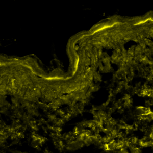
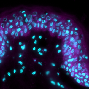
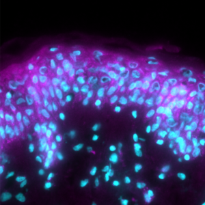
Test: Mitofusin-2 (Mfn2) expression
Interest: Mitofusin-2 controls mitochondrial fusion, the process by which mitochondria merge to maintain a healthy, connected network. Its decline under stress reflects loss of structural integrity.
Method: Immunofluorescence imaging
Model: Ex vivo human skin, UV stress model
Interpretation of results: UV stress reduced Mfn2 signal versus control. Product pretreatment partially restored expression, indicating protection of the mitochondrial fusion machinery.

CELLULAR HEALTH LEVEL
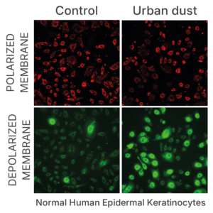
Test: Mitochondrial membrane potential
Interest: Membrane potential is one of the most reliable indicators of mitochondrial health. Depolarisation signals functional impairment and precedes cell damage.
Method: JC-1 ratiometric fluorescence (red/green ratio)
Model: Normal human epidermal keratinocytes (NHEK), urban dust stress model
Interpretation of results: Urban dust significantly reduced the red/green JC-1 fluorescence ratio versus control, indicating membrane depolarisation and mitochondrial impairment.

StressProduct + stress

MITOCHONDRIAL MASS & METABOLIC LEVEL
Test: Citrate synthase expression
Interest: Citrate synthase is a rate-limiting enzyme of the TCA cycle (the cell’s central energy cycle) and a recognised proxy for mitochondrial mass. Its level reads out both metabolic capacity and the size of the functional mitochondrial pool.
Method: Enzymatic expression quantification
Model: Oxidative stress model
Interpretation of results: Oxidative stress reduced citrate synthase to 57 ± 9% of control. Product pretreatment restored it to 98 ± 2% — near-complete preservation of mitochondrial mass under stress. The numeric restoration gives a defensible, quantified protection readout.

FUNCTIONAL ENERGY LEVEL
Test: Mitochondrial respiration (Seahorse OCR)
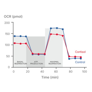
Interest: Oxygen consumption rate is the most direct functional readout of mitochondrial respiration. A single run resolves three phases — basal respiration, ATP-linked respiration, and maximal respiratory capacity — so you measure energy output itself, not a static marker.
Method: Seahorse XF real-time oxygen consumption rate (OCR) profiling
Model: Normal human epidermal keratinocytes (NHEK), cortisol-induced metabolic stress
Interpretation of results: Cortisol reduced OCR across all three phases versus control, confirming impaired capacity to produce ATP. The assay quantifies whether an active restores respiration under stress — a functional efficacy readout that goes straight into a claims file.

4. Workflow: From Sample to Report

5. Who We Work With

  • Cosmetics brands: Cosmetics and dermo-cosmetics brands substantiating skin longevity or “skin energizing” claims under EU Cosmetics Regulation 1223/2009
  • Ingredient suppliers: Ingredient suppliers developing actives targeting mitochondrial fitness, cellular energy, or anti-aging efficacy
  • R&D teams: R&D and formulation teams screening compounds for functional effect on mitochondrial respiration or membrane potential in primary skin cells
  • Contract labs: Contract formulation labs needing regulatory-ready in vitro data packages before client submission or product launch

6. Why QIMA Life Sciences for Mitochondrial Dysfunction Assays

  • Dedicated team of cell biologists and biochemists specialised in skin aging research
  • Integrated assay platform covering the full spectrum of mitochondrial readouts from functional to structural
  • Customised study design aligned with your specific claim objectives and regulatory requirements
  • End-to-end continuity: the same scientific team supports you from in vitro screening to clinical validation
  • Data packages structured for claim dossier integration and regulatory submission
  • From biological readout to consumer claim: we structure the evidence chain so your product story holds at every level from in vitro mechanism to perceived age improvement

Get the full scientific overview of mitochondrial dysfunction, assay protocols, and data highlights.

mitrochondrial dysfunction flyer miniature

Frequently Asked Questions

Primary human dermal fibroblasts (NHDF) and epidermal keratinocytes (NHEK) are the primary cell types used, sourced from a bank of over 300 donors covering a range of ages, sexes, and anatomical sites. 3D reconstructed skin models and ex vivo human skin explants are also compatible for structural and imaging readouts. Each assay is optimised per cell type and study objective.

The Seahorse XF assay measures oxygen consumption rate (OCR) in real time, providing a quantitative readout of mitochondrial respiration across 3 key phases: basal respiration, ATP-linked respiration, and maximal respiratory capacity. These parameters directly support claims related to skin energising, mitochondrial fitness, and cellular vitality under EU Cosmetics Regulation 1223/2009.

Yes. Our modular study design supports parallel testing of multiple hallmarks within a single protocol. Mitochondrial dysfunction assays can be combined with senescence markers (SA-β-gal, p16, p21), inflammaging readouts (IL-6, IL-8, TNF-α), oxidative stress panels, or telomere attrition assays.

Yes. QIMA Life Sciences provides raw data, statistical analysis, and a narrative study report structured to support claim substantiation files under EU Cosmetics Regulation 1223/2009.

Both. The mitochondrial assay portfolio applies to 2D in vitro models (primary keratinocytes and fibroblasts) and to ex vivo human skin explants, including a 3D ex vivo model of human skin mimicking intrinsic aging. Model selection is guided by the study objective and the claims to be substantiated. Contact our team for more info.

At QIMA Life Sciences, we specialize in providing comprehensive solutions for studying skin aging and longevity through validated models at both preclinical and clinical levels.

Our dedication to scientific innovation drives us to develop cutting-edge models and approaches, staying aligned with the latest advancements in longevity research.

Explore Other Resources

Image of a computer displaying a webpage called "Assays to study the hallmarks of skin aging"
Image of a computer displaying a webpage called "Skin Longevity & Aging"
Image of a computer displaying a blog article called "A new era in skin aging"
Graduation-cap Graduation-cap

Related Publications

This field is for validation purposes and should be left unchanged.

Hello! How can we help you?

Name(Required)

By contacting QIMA Life Sciences you agree to our privacy policy and terms and conditions.