


What is slide scanning?
At a time of biological data computerization, slide scanning (virtual slides) has become a major tool for cosmetics research.
Virtual microscopy consists in scanning and digitizing a selected field in order to obtain a global view of the biological materials that are being observed. In addition to the high resolution viewing of the slides, this method allows us to move on the slide, to adjust the brightness and to zoom in areas of interest.
You can virtually view this image (in jpg or tiff format) from any computer, as you would on a microscope. We also benefit from a digital storage of these slides for validation and archiving purposes.
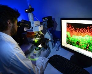
Bioalternatives provides you with the ability to semi-automatically digitize your slides, by using a Nikon Eclipse Ci straight microscope equipped with a motorized X-Y-Z stage (autofocus). This system enables the capture of « big images » at x10, x20 or x40 magnifications (contact us).
This system is suitable for histology, immunohistochemistry and immunofluorescence techniques.
Examples of slide scanning
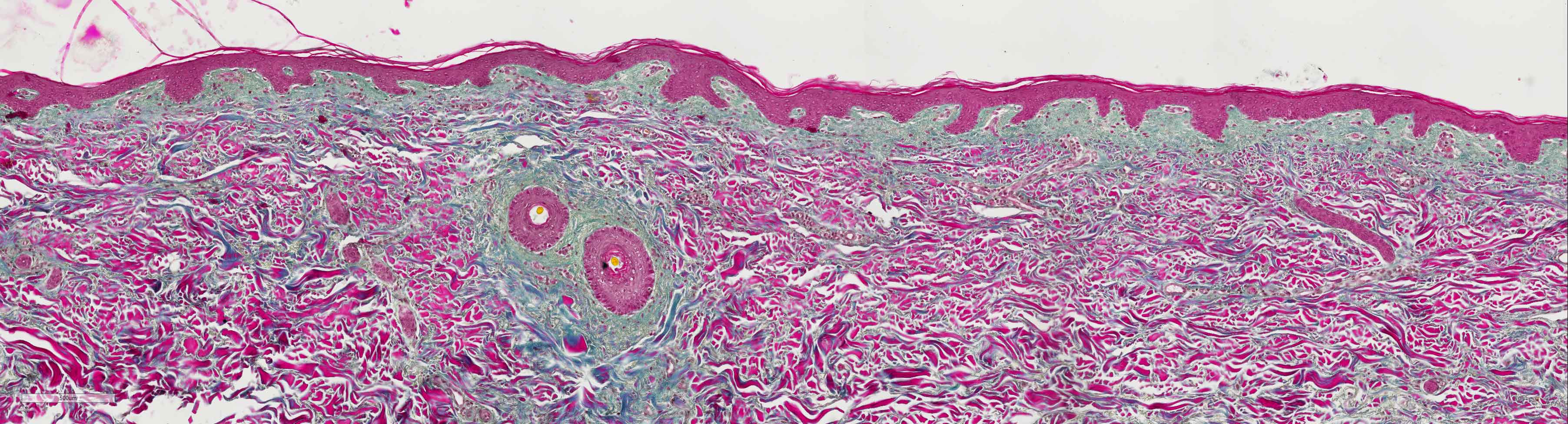
Scanning of explant section of colored human skin (HES) x40
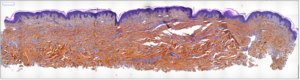 Whole non-zoomed scanned image
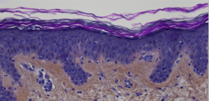 Zoomed scanned image
Scanning of explant section of fluorescence-labeled human skin x40
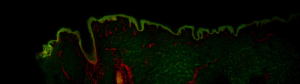 Whole non-zoomed scanned image
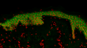 Zoomed scanned image
- CDH1 or E-cadherin in green (membrane labeling)
- Propidium iodide in red (nucleus)


