

Blog

We are passionate about what we do

and we want to share that passion with you.

TECHNICAL FOCUS:

Immunocompetent Skin Model with Integrated Dendritic Cells

Many reconstructed skin models replicate the skin’s structural barrier but not its immune function. This can limit their utility in evaluating inflammatory skin responses or topical product tolerance, and in mechanistic research on topics such as allergic contact dermatitis.

Our immunocompetent full thickness skin (FTSK) model closes that gap by integrating monocyte-derived dendritic cells (DCs) directly into the dermal compartment. Below is the imaging and functional data showing how the integration of DCs potentiates cytokine responses, along with what the model can be used for.

Key takeaway:

Our immunocompetent FTSK-DC model integrates functional monocyte-derived dendritic cells into a full thickness human skin, confirmed by immunofluorescence. This enables a pharmacologically reversible cytokine response to LPS challenge that the DC-free model does not produce.

This model has uses in topical immunotoxicity screening, allergic contact dermatitis research, and inflammatory skin response evaluation, among other applications, and particularly suits programs with reduced reliance on animal models, making it a strong fit for new approach methodology (NAM)-aligned development strategies.

Immunocompetent FTSK Model Architecture

The model is a reconstructed full thickness human skin with monocyte-derived DCs seeded into the dermal compartment alongside keratinocytes and fibroblasts. Immunofluorescence staining for HLA-DR and CD209 (DC-SIGN) confirms DC integration and distribution throughout the tissue.

Need a different

configuration?
This model sits within a broader custom skin tissue platform, including barrier-defect models, cytokine-induced disease states, stressed skin phenotypes, host-microbe co-culture systems, and much more. Access our brochure above for more information.
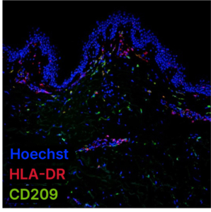
Immunofluorescence staining of healthy human skin ex vivo.
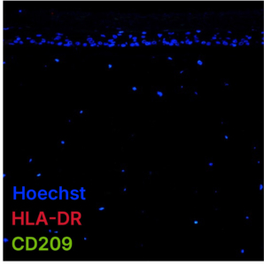
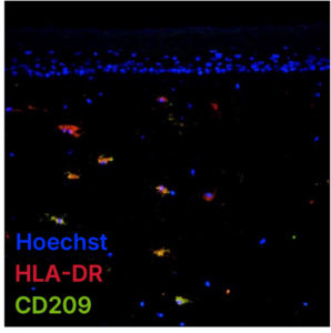
FTSKFTSK-DC

Comparison between the standard FTSK model and the immunocompetent FTSK-DC model, showing the integration of DCs.
Cytokine Response of the Immunocompetent Skin Model

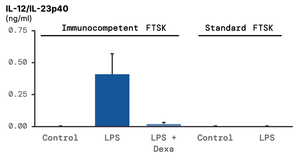
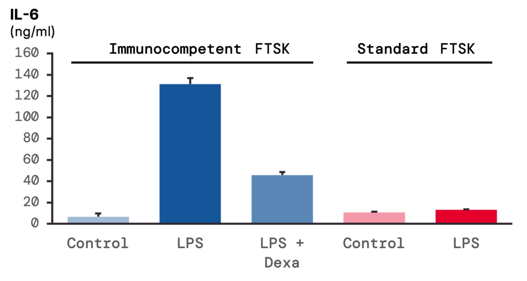
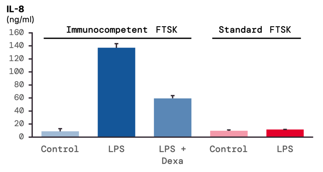
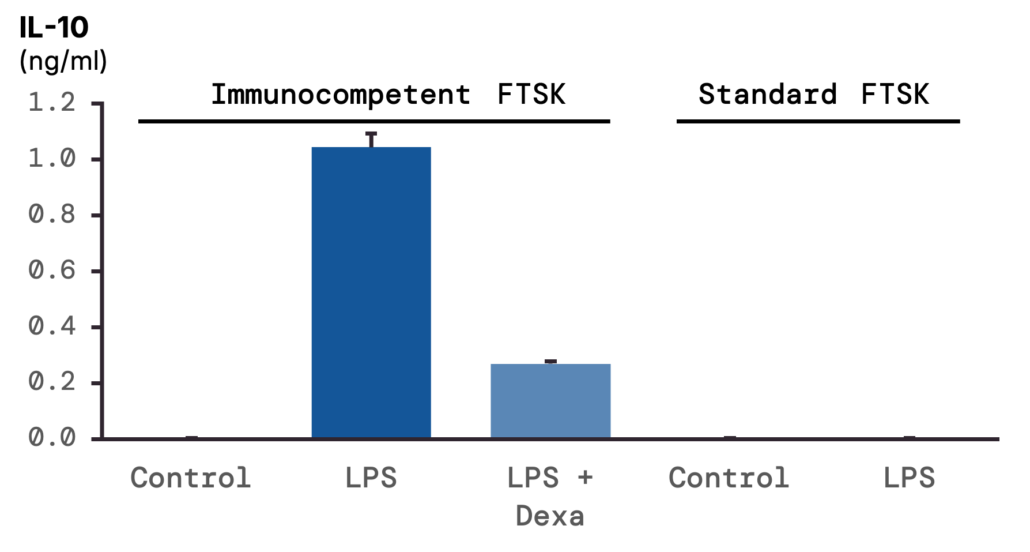
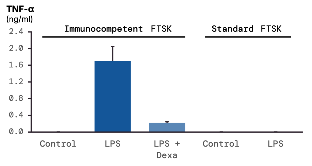
Following systemic LPS stimulation, the FTSK-DC model releases a distinct cytokine array that the DC-free model largely does not. In side-by-side testing under identical LPS exposure:

  • TNF-α, IL-12/IL-23p40, and IL-10 rise to clearly quantifiable levels in the DC-containing model, while remaining below the assay’s detection limit in the DC-free model under the same conditions.
  • IL-6 and IL-8 increase roughly tenfold in the FTSK-DC model relative to the DC-free model under the same stimulus.

The model was also challenged with LPS in the presence of dexamethasone, a reference anti-inflammatory corticosteroid, at 1 µM. Relative to LPS alone, co-treatment reduced:

  • TNF-α by roughly 85%
  • IL-12/IL-23p40 by roughly 95%
  • IL-6 by roughly 65%
  • IL-8 by roughly 55%

IL-6 and IL-8 release was notably high in this model in response to LPS challenge. Other cell types present in the model, particularly fibroblasts, may also respond and contribute to the release of these cytokines, in addition to the integrated dendritic cells.

Applications of the Immunocompetent FTSK

The combination of structural integration and a functional, intervenable cytokine output makes this model suited to:

  • Topical immunotoxicity and immunomodulation screening for drug candidates and formulations.
  • Tolerance assessment work where a DC-free model may under-report inflammatory potential.
  • Mechanistic studies of dendritic cell activation and suppression in a tissue-relevant context, ahead of in vivo work.
  • Comparator testing against DC-free models to isolate the contribution of the immune compartment specifically.

Reconstructed skin models have established OECD test guidelines for corrosion and irritation testing applications, and dendritic cell activation is an endpoint increasingly used as a non-animal readout for adverse outcomes such as skin sensitization. This model can offer an exploratory indication of both outcomes, making it a valuable tool for NAM-aligned product development.

The model forms part of our immunology toolkit, and can be adapted for donor selection, alternative stimuli, or compound panels as part of a custom study design. Our future perspectives for immunocompetent skin modelling include the optimisation of topical stimuli to induce DC activation and cytokine release, and the progressive integration of additional immune populations, such as monocyte-derived Langerhans cells, macrophages, and/or T cells.

Written by:

Callum O’Kane, PhD

Marketing Project Leader – Pharmacology

Edited by:

Sabrina Hoefling, PhD

Marketing Project Leader – Dermatology

Interested in running your compound or formulation through this model?

This field is for validation purposes and should be left unchanged.

Hello! How can we help you?

Name(Required)

By contacting QIMA Life Sciences you agree to our privacy policy and terms and conditions.