
Related Publications
SELECTED PUBLICATIONS:
Pemphigoid disease model systems for clinical translation
>> Check the full list HERE.
Conference Contributions
We share our research on dermatology and trichology through international conferences and publications, contributing to the advancement of research in these fields.
>> Check our past contributions HERE.
What we help you achieve
- Evaluate candidate therapeutics against key pemphigoid effector mechanisms (neutrophil activation and complement activation).
- Generate mechanism-focused preclinical evidence to support lead selection and early proof-of-concept.
- Benchmark candidates against current therapies within a controlled assay framework.
- Reduce reliance on animal studies by filtering and prioritizing compounds using human skin and donor-derived cells.
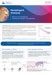
Ex Vivo Models for Pemphigoid Diseases
- ROS-release assay: Measures reactive oxygen species from human neutrophils upon immune-complex activation. Ideal for screening anti-inflammatory small molecules.
- Cryosection assay: Human skin cryosections incubated with patient or recombinant IgG to quantify dermal–epidermal split formation. Validates mechanism in a tissue context.
- Complement fixation assay: Fluorescence-labeled C3/C5 antibody binding to immune complexes on skin sections. Perfect for pipeline testing of complement inhibitors.

Pemphigoid Disease Models Available Now
IMMUNE COMPLEX-INDUCED NEUTROPHIL ACTIVATION
Test: ROS-release assay (AUC readout)
What it measures: Reactive oxygen species (ROS) release from immune complex–activated neutrophils.
Assay principle:
- Immune complex (IC) formed on a plate using an autoantibody and its corresponding antigen
- Neutrophils isolated from healthy blood donors are added
- ROS-driven luminol chemiluminescence is measured
- Neutrophil activation is quantified from the area under the luminescence curve (AUC)
Use case: Screening and ranking compounds based on their ability to reduce neutrophil activation.
NEUTROPHIL-DRIVEN DERMAL-EPIDERMAL JUNCTION DAMAGE
Test: Cryosection split assay
What it measures: Split formation at the dermal–epidermal junction (DEJ) in human skin sections.
Assay principle:
- Incubate healthy human skin sections with autoantibodies
- Incubate with neutrophils isolated from healthy blood donors
- Split formation is induced at the DEJ
- Readout is the percentage of split formation
Use case: Tissue-level confirmation of functional protection beyond cell-based readouts.
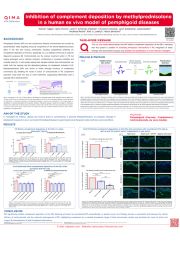
COMPLEMENT BINDING AND ACTIVATION AT THE DEJ
What it measures: Complement deposition along the DEJ via autoantibody-mediated fixation.
Assay principle:
- Incubate healthy human skin sections with autoantibodies, then with normal human serum (complement source)
- Complement components are fixed to the DEJ
- Detection is performed using a fluorescence-labelled anti-complement C3c antibody
- Readout is fluorescence intensity along the DEJ (inhibition assessed as reduced signal)
Use case: Testing complement-pathway intervention strategies in a human tissue context.


At QIMA Life Sciences, we are committed to staying at the forefront of dermatology research by developing innovative approaches.
We offer smart solutions for studying pemphigus and pemphigoid diseases using validated models, making us the perfect partner for your research.
Explore Our Models & Assays in Our Flyer and Poster