- Development:
- staining
- immunolabeling (research and validation of antibodies)
- co-labeling
- Preparation of slices
- Images, image mosaic, slide scanning, TEM/SEM
- Quantification / image analysis (thickness, intensity, counts, etc.)

HISTOLOGY
IMMUNOHISTOCHEMISTRY
IMMUNOFLUORESCENCE
KEY FIGURES
staining methods
validated antibodies for IHC/IF
analyzed samples per year
pictures per year
Read more
Sample & slide preparation
 The sample and slide preparation is a crucial step for histological or cytological observation. It is crucial to highlight what needs to be observed and to « immobilize » the sample at a particular point in time and with characteristics close to those of its living state. Bioaternatives sets up sample and slide preparation protocols that guarantee an observation which is as close as possible to the actual living tissues…
Histological staining

At Bioalternatives, we offer more than thirty standard (topographic) histological and special (descriptive) staining techniques.
These staining processes are carried out after a thorough preparation of samples and slides.
With routine standard staining techniques, tissue morphology (structure, organization, dimension / nucleus, cytoplasm, collagen fibers) can be examined and tissue integrity or …
Immunolabeling: IF/IHC

Bioalternatives offers immunolabeling services in order to detect the cell and tissue components in a tissue section. Proteins of interest (antigens) are detected by using polyclonal or monoclonal antibodies specifically directed against the target proteins.
The detection methods directly depend on the type of marker that is used. Our laboratory uses special in situ detection techniques: immunofluorescence and immunohistochemistry.
Electron microscopy service
Do you wish to illustrate your studies, support your quantitative results and optimize your visual marketing supports with striking high quality images?
Bioalternatives provides you with a full service of sample analysis and TEM or SEM observation and imaging…
Slide scanning service
Do you wish to illustrate your studies, support your quantitative results and enrich your marketing materials with an innovative digital approach?
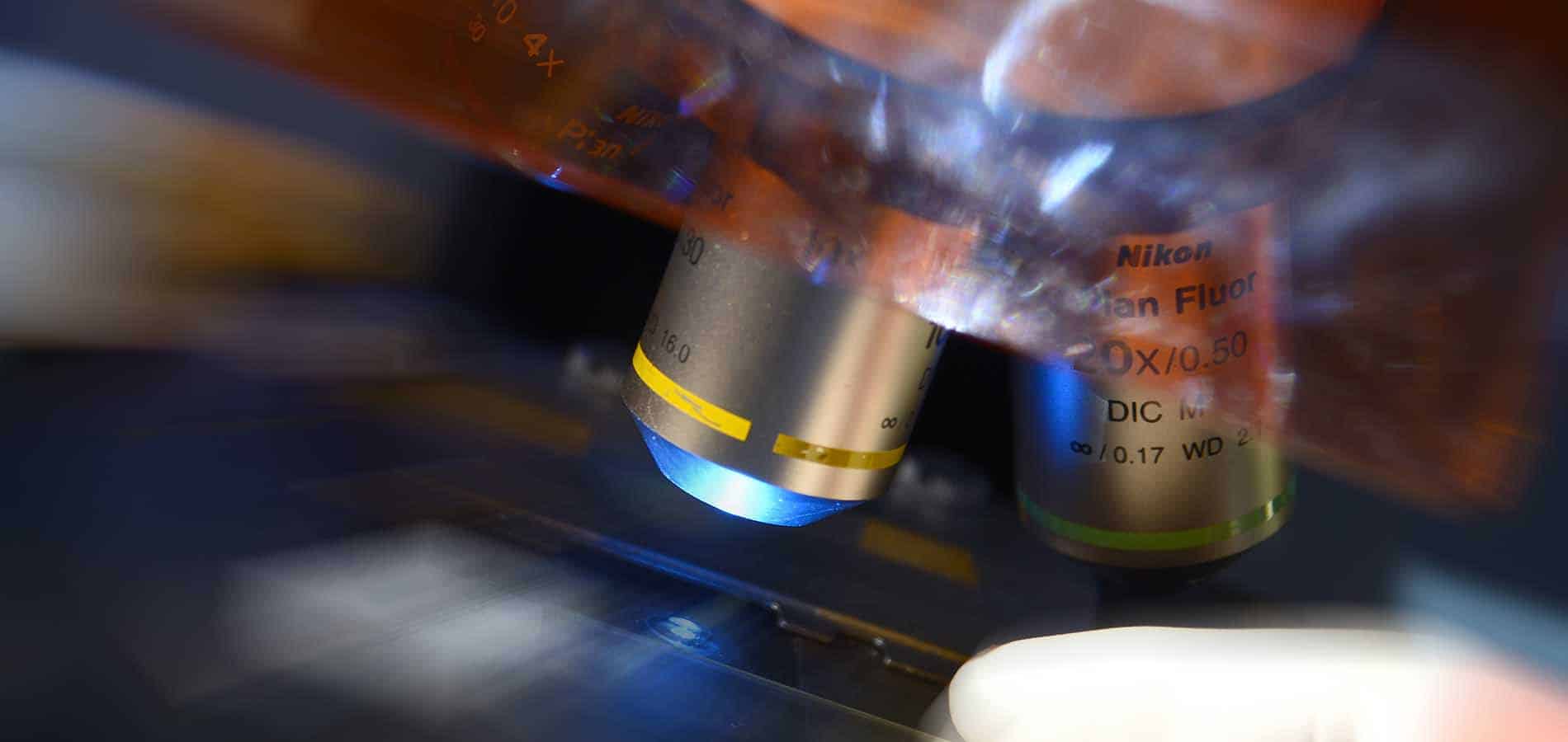
Bioalternatives provides you with the ability to semi-automatically digitize your slides, by using a Nikon Eclipse Ci straight microscope equipped with a motorized X-Y-Z stage (autofocus). This system enables the capture of « big images » at x10, x20 or x40 magnifications.
This system is suitable for histology, immunohistochemistry and immunofluorescence techniques.
Cosmetics & Personal Care
Pharma & Biotech
Search
France
QIMA Bioalternatives:
+33 (0)5 49 36 11 37 (Gençay)
+33 (0)5 61 28 71 60 (Labège)
QIMA Newtone:
+33 (0)4 72 69 83 20
QIMA Skinpharma:
+33 (0)4 92 03 62 40
Germany
QIMA Monasterium:
+49 (0)251 93264458


