

Mitochondrial Dysfunction Assays for Skin Longevity Claims
The cosmetics market is converging on longevity science. Brands that demonstrate an ingredient’s effect on cellular energy metabolism, rather than surface hydration or wrinkle reduction alone, are staking out a scientifically differentiated position. That requires purpose-built models and quantifiable readouts.
At QIMA Life Sciences, we design mitochondrial studies around your specific claim objective: from early ingredient screening to multi-readout evidence packages structured for regulatory claim dossiers.
On this page:
- Key mechanisms linking mitochondrial dysfunction to skin aging
- QIMA Life Sciences’ quantitative assay platform: Seahorse OCR, JC-1, MTT, NAD+/NADH, MitoTracker, Mitofusin-2
- Workflow from sample preparation to claim-ready report
- Why QIMA Life Sciences
Why Mitochondrial Dysfunction Matters for Skin Aging
Mitochondria regulate ATP production, ROS balance, and the initiation of apoptosis. In skin cells, age-related mitochondrial decline creates a cascade of bioenergetic deficits that drive structural aging long before visible signs appear:
- Reduced oxidative phosphorylation lowers ATP output, compromising keratinocyte turnover and extracellular matrix maintenance.
- Elevated ROS production, from UV exposure and internal mitochondrial sources, induces oxidative damage to proteins, lipids, and mtDNA, activating senescence pathways in dermal cells.
- Progressive loss of membrane potential, slower biogenesis, and a shift in the fusion/fission balance compound the energy deficit over time.
These disruptions manifest as fine lines, reduced elasticity, impaired barrier function, and altered skin tone. Demonstrating that your ingredient acts upstream on the energy deficit driving these changes is the scientific foundation of a credible skin longevity claim.
1. Mechanisms of Mitochondrial Dysfunction in Skin Cells
Impaired oxidative phosphorylation
Reduced ATP production compromises keratinocyte turnover and extracellular matrix maintenance, directly affecting the skin’s capacity to repair and regenerate. Evaluating this mechanism requires real-time measurement of mitochondrial respiration, not endpoint markers.
Elevated mitochondrial ROS
An imbalance between ROS production and antioxidant defenses induces oxidative damage to proteins, lipids, and DNA, activating senescence pathways in dermal fibroblasts and keratinocytes. ROS quantification in relevant cell models provides a mechanistic readout usable directly in a claims file.
Mitochondrial structural and functional decline
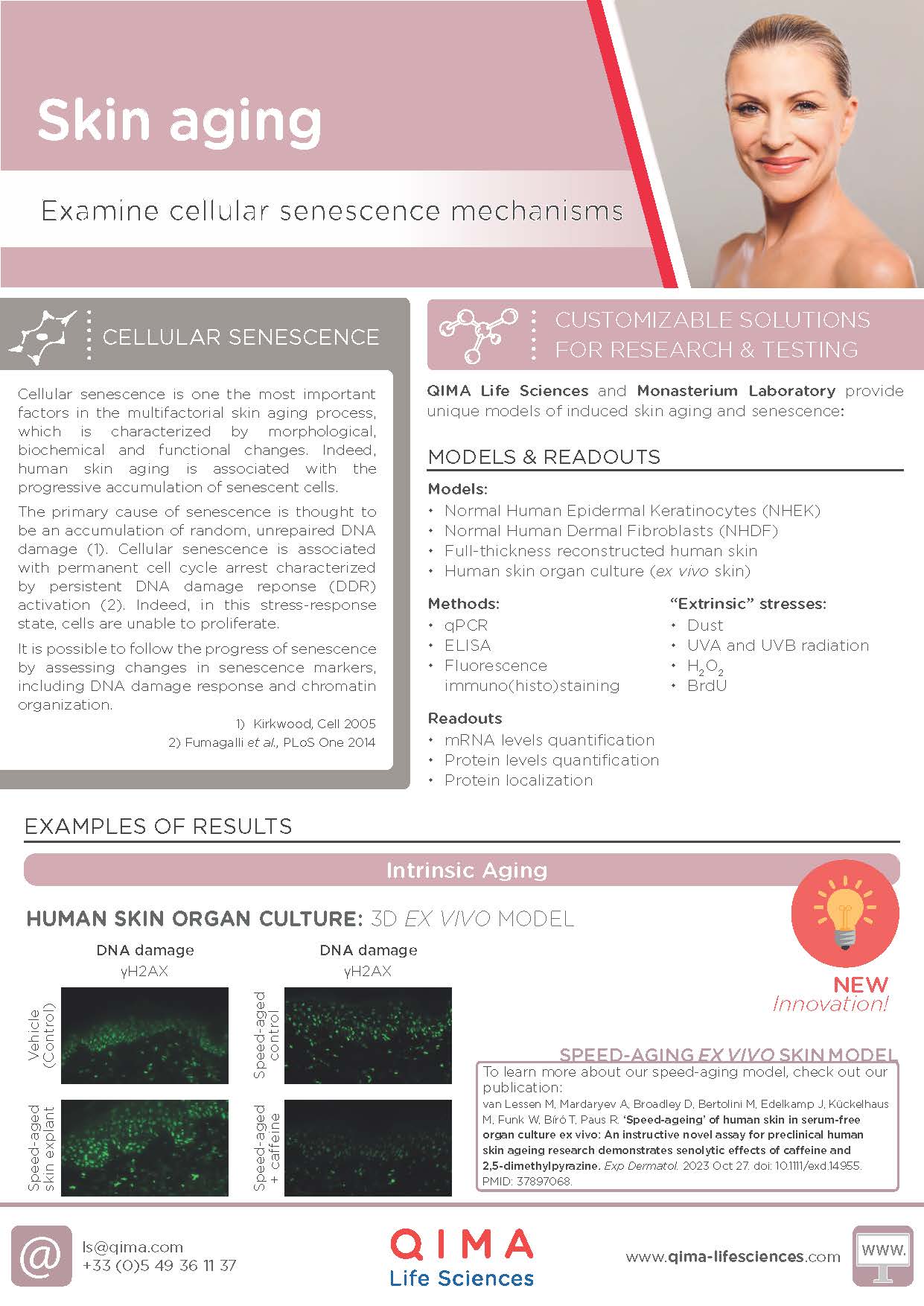
With age, mitochondrial integrity deteriorates: membrane potential decreases, biogenesis slows, and the fusion/fission balance shifts. Fluorescence-based structural imaging in ex vivo human skin, including Mitofusin-2 (Mfn2) quantification as a marker of fusion activity, captures these changes at the tissue level.
2. Consequences for Skin Aging
These bioenergetic changes translate into observable skin aging outcomes:
- Barrier dysfunction elevates transepidermal water loss (TEWL) and increases sensitivity
- Keratinocyte and fibroblast senescence reduces regenerative capacity and impairs repair
- Chronic low-grade oxidative stress sustains inflammaging, accelerating collagen degradation
Substantiating a claim at this level means showing your ingredient shifts a functional parameter, not just co-localising with a marker. That is the standard QIMA Life Sciences applies when structuring a study.