


Share this Technical Focus
Key takeaway:
The replicative senescence fibroblast model provides a robust in vitro platform for assessing the impact of cosmetic actives on intrinsic skin aging mechanisms.
Understanding the biological mechanisms behind intrinsic skin aging is essential for developing innovative anti-aging interventions.
Our team has established a fibroblast senescence model based on the Hayflick limit, which accurately mimics age-related cellular exhaustion (Hayflick L, 1961).
Download our flyer on
Cellular Senescence

Aging Biomarkers and Cellular Alterations
With successive cell divisions, dermal fibroblasts progressively lose their proliferative capacity and enter a senescent state, exhibiting:
- Morphological changes: Enlarged, flattened cells with altered cytoskeletal organization.
- Senescence markers: Increased expression of proteins regulating the cell cycle, such as p16, which leads to irreversible cell cycle arrest, along with a marked decrease in Ki67, a key proliferation marker.
- Extracellular matrix (ECM) remodeling: Decreased synthesis of collagen, hyaluronic acid, glycosaminoglycans, and elastin, along with upregulated MMPs (MMP-1, MMP-3), leading to matrix degradation.
- Oxidative stress and SASP-driven inflammation: Senescent fibroblasts develop a SASP, which sustains chronic low-grade inflammation (inflammaging) and exacerbates oxidative stress.
Download our brochure on
Skin Longevity
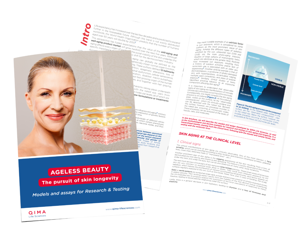
Using this well-characterized model, our team enables targeted efficacy testing, supporting the development of next-generation cosmetic actives designed to promote skin longevity by preserving cellular function, extracellular matrix integrity, and resilience over time.
ECM Remodeling
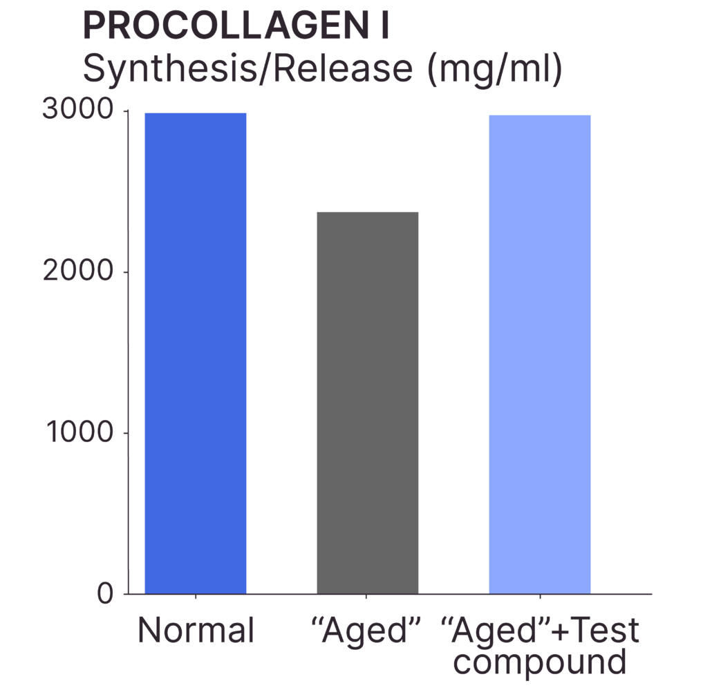
Test: Procollagen I Synthesis
Method: ELISA
Observation: The addition of the test compound prevents the decline in procollagen I synthesis observed in senescent fibroblasts.
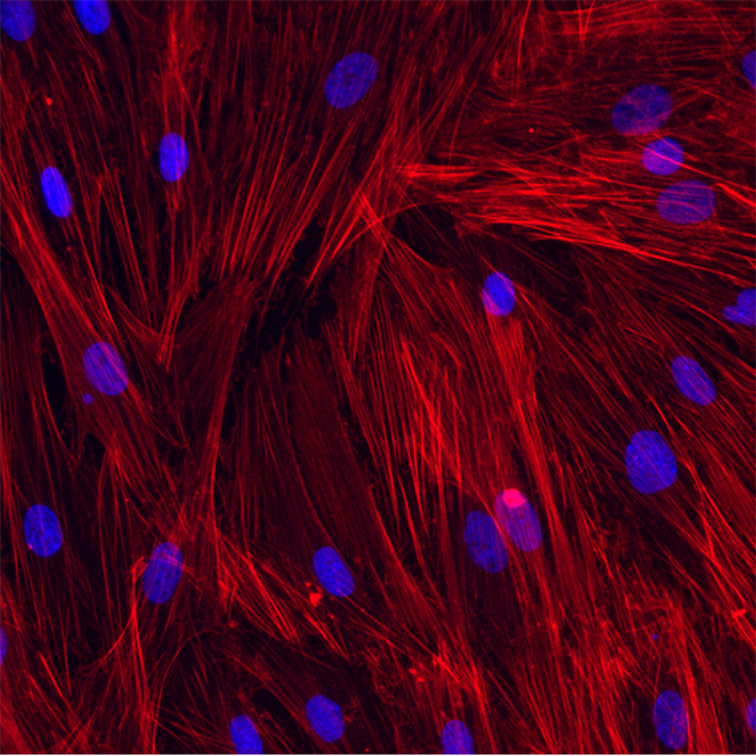
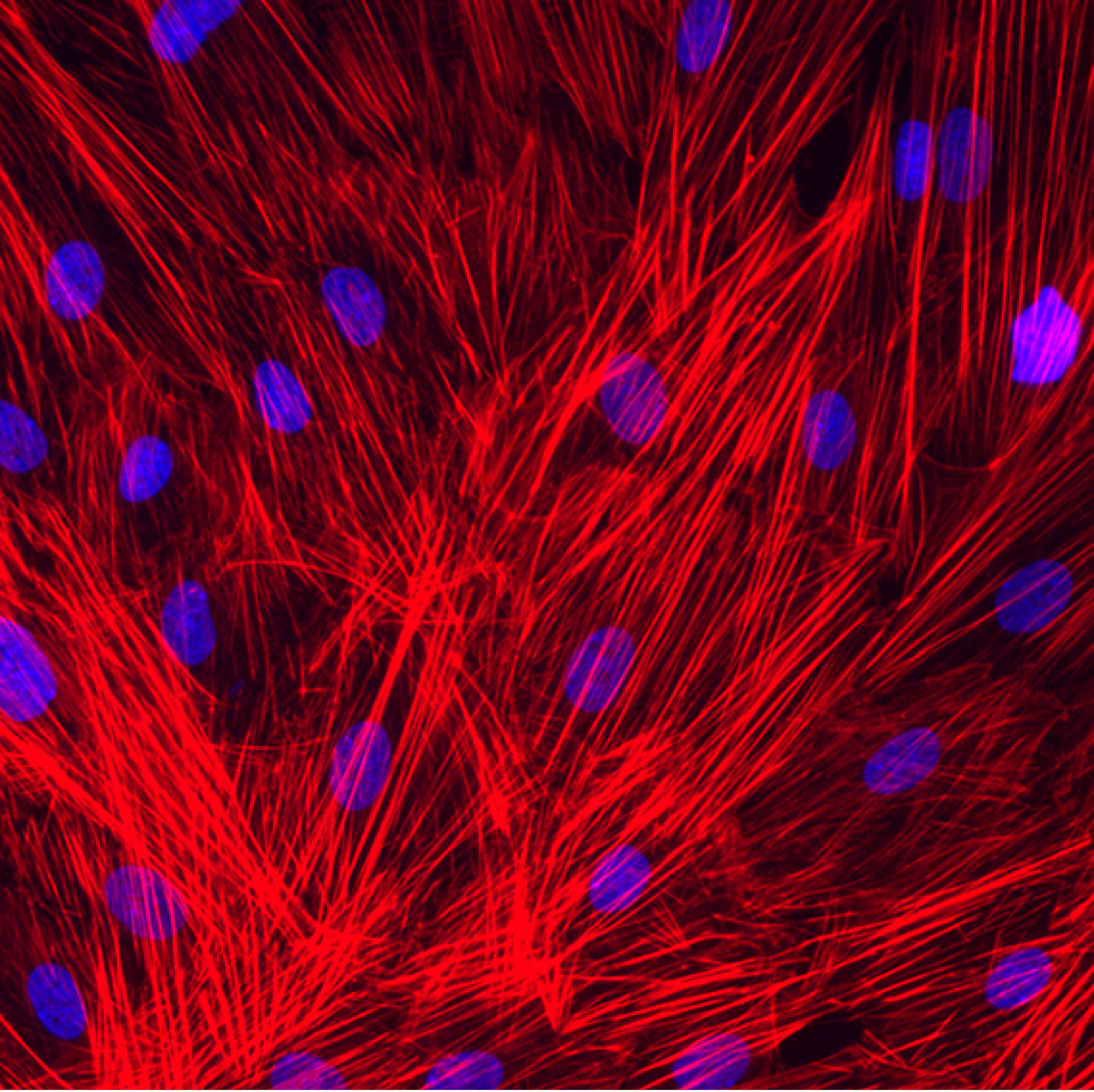
Cytoskeletal organization
Test: Actin Expression
Method: Immunofluorescence (actin: red)
Observation: Actin expression is reduced in senescent fibroblasts, which leads to defects in cytoskeletal organization. Decrease in actin expression is prevented by the addition of the test compound.
Migration capacity
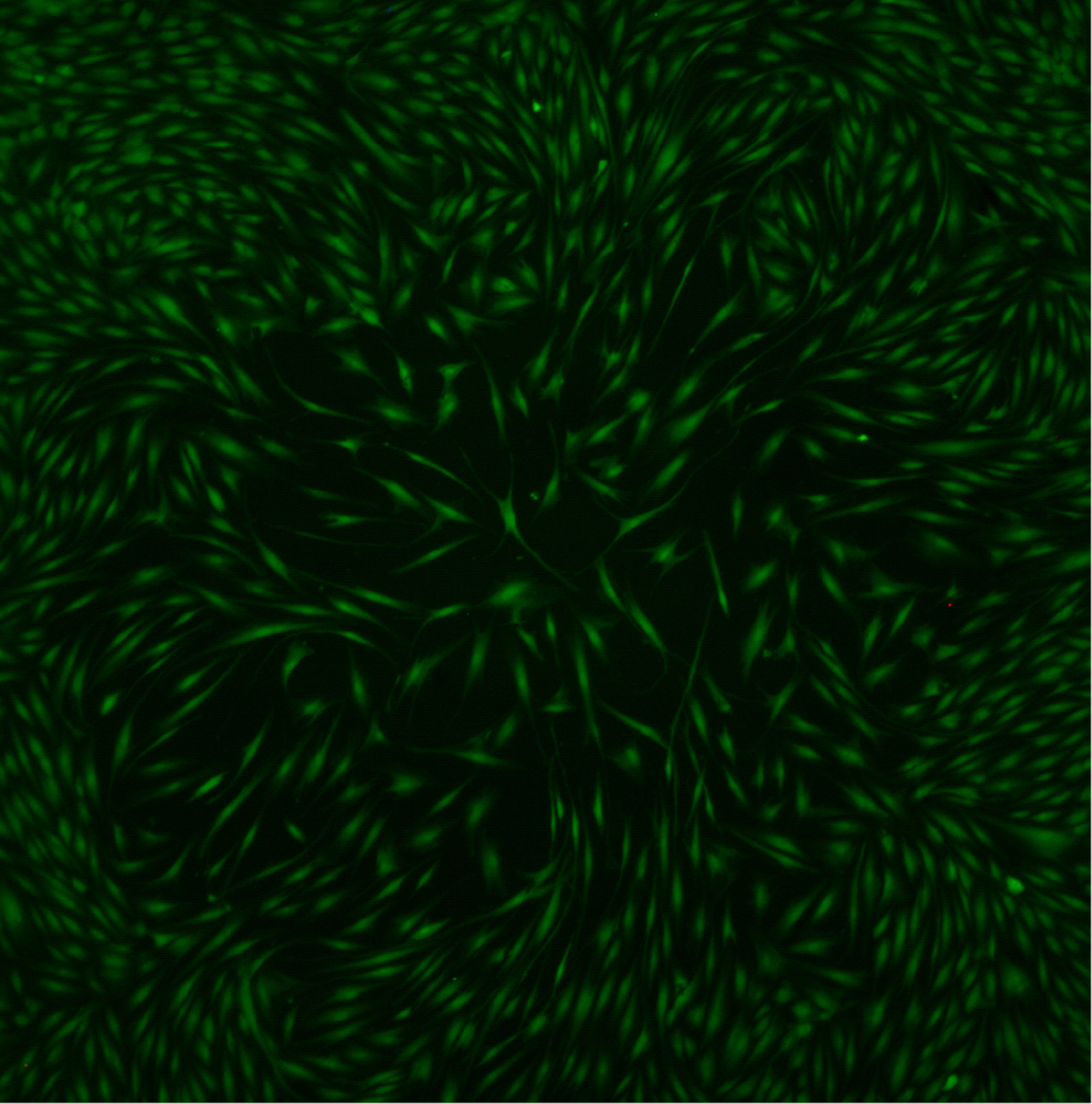
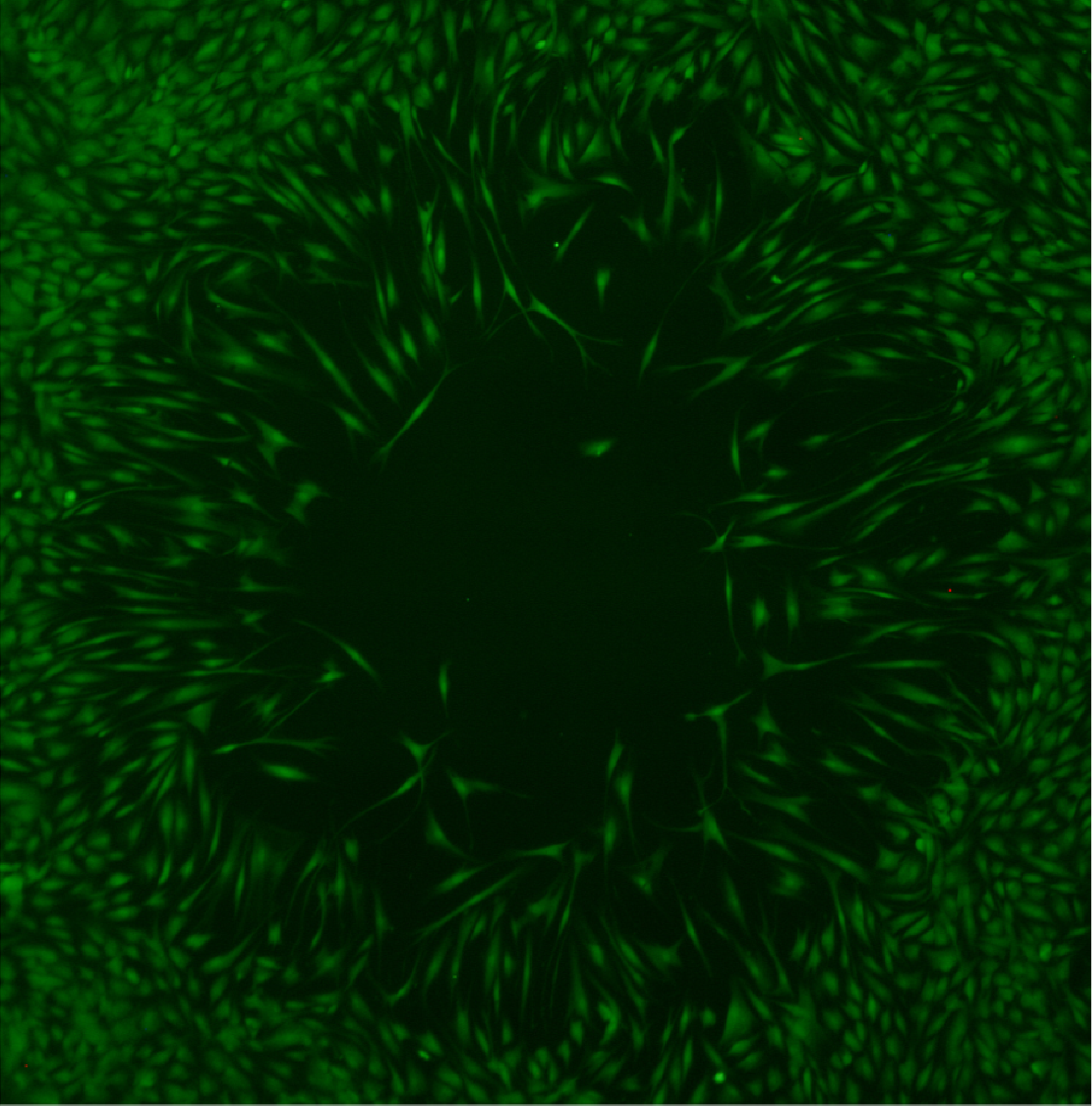
Test: Fibroblast Migration
Method: Immunofluorescence
Observation: Senescent fibroblasts exhibit reduced migratory capacity, leading to a prolonged time to achieve complete closure of a cell-free area. Results shown correspond to the same time point.
Dermal Contractility
Test: Dermal Contraction
Method: Surface Area Measurement
Observation: Senescent fibroblasts exhibit reduced contractile capacity.
Read related Technical Focus

Written by:
Mara Carloni, PhD
Scientific Communications & Marketing Project Leader