Related Publications
SELECTED PUBLICATIONS ON HIDRADENITIS SUPPURATIVA
Exploring the human hair follicule microbiome
>> Check the full list HERE.
Conference Contributions
We share our research on dermatology and trichology through international conferences and publications, contributing to the advancement of research in these fields.
- Exploring initial events in Hidradenitis Suppurativa pathogenesis by utilizing the hair follicle ex vivo model (EHRS 2025)
- Deciphering Secukinumab’s mechanism in reducing hidradenitis suppurativa inflammation: Insights from diseased skin organ culture (AAD 2025)
- Exploring the potential of selective BET inhibition as a treatment for Hidradenitis suppurativa (EHSF 2025)
>> Check our past contributions HERE.
Preclinical Research Solutions for Hidradenitis Suppurativa
IN VITRO MODELS
- Outer Rooth Sheath Keratinocytes
- PBMCs from healthy donors or HS patients
- Skin-derived immune cells from healthy donors or HS patients
EX VIVO MODELS
- Occipital healthy human hair follicles ex vivo
- Induction of HS phenotype in occipital healthy human hair follicles ex vivo
- Co-culture of human hair follicles with autologous blood- or skin-derived immune cells
- Healthy human (scalp) skin ex vivo
- Organ culture of peri-lesional, nodule-containing, or fistula-containing lesional biopsies from HS patients
We can help you evaluate the following readouts
Cytokine & Chemokine Release
Spatial Proteomics (up to 40 markers)
Spatial Transcriptomics
RNAseq
scRNAseq
…among many others.
Clinical Research Solutions for Hidradenitis Suppurativa
BIOANALYSIS OF CLINICAL SAMPLES
- Sample collection (skin biopsies)
- Analysis and quantification of cellular components (proteins, lipids) via analytical chemistry
- HS biomarker analysis
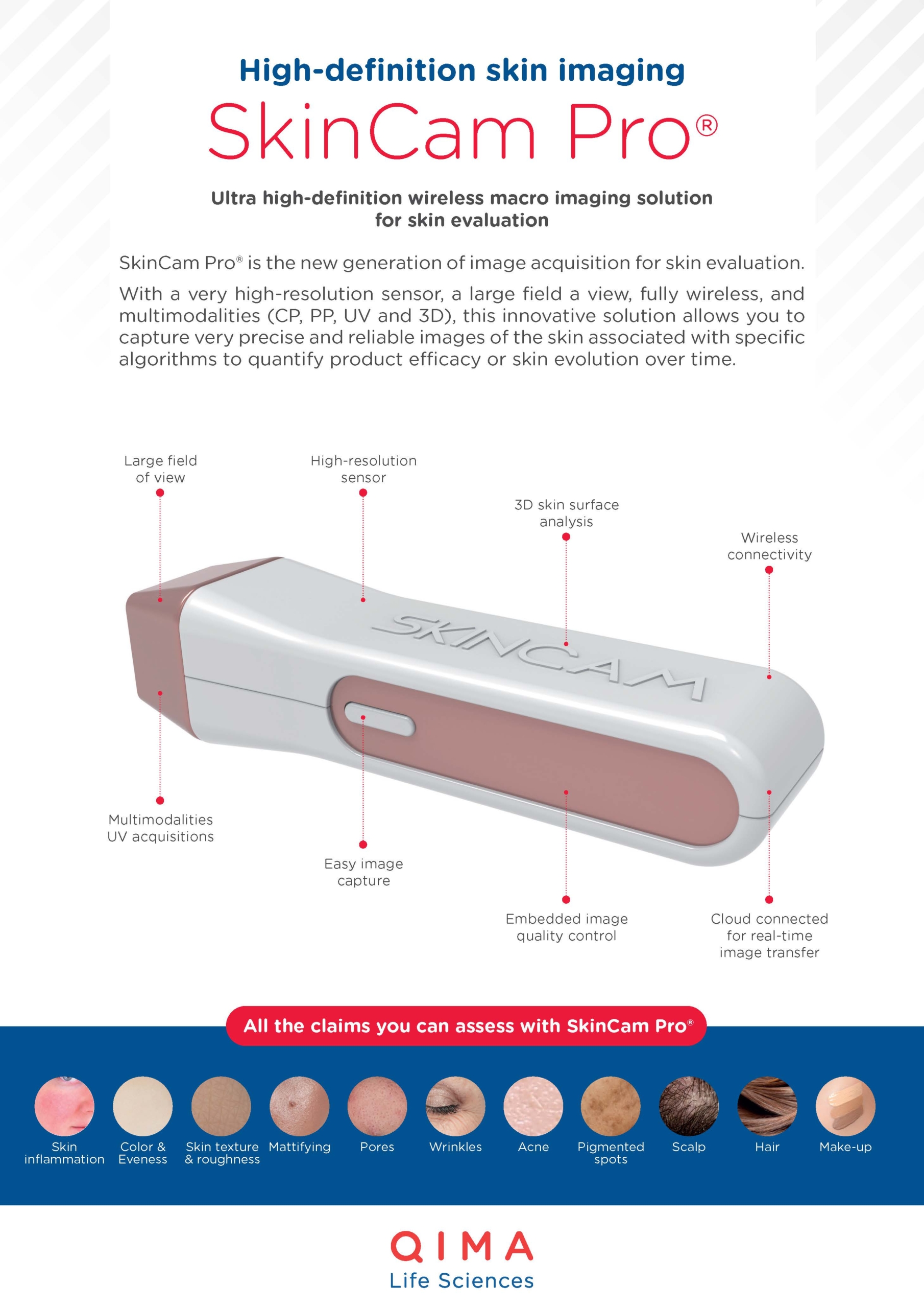
CLINICAL IMAGING
- Image capture
- Tracking of HS lesions
CLINICAL TRIALS
- Clinical study implementation
- Clinical study performance
- Data management
- Data analysis
Hidradenitis Suppurativa Study Examples
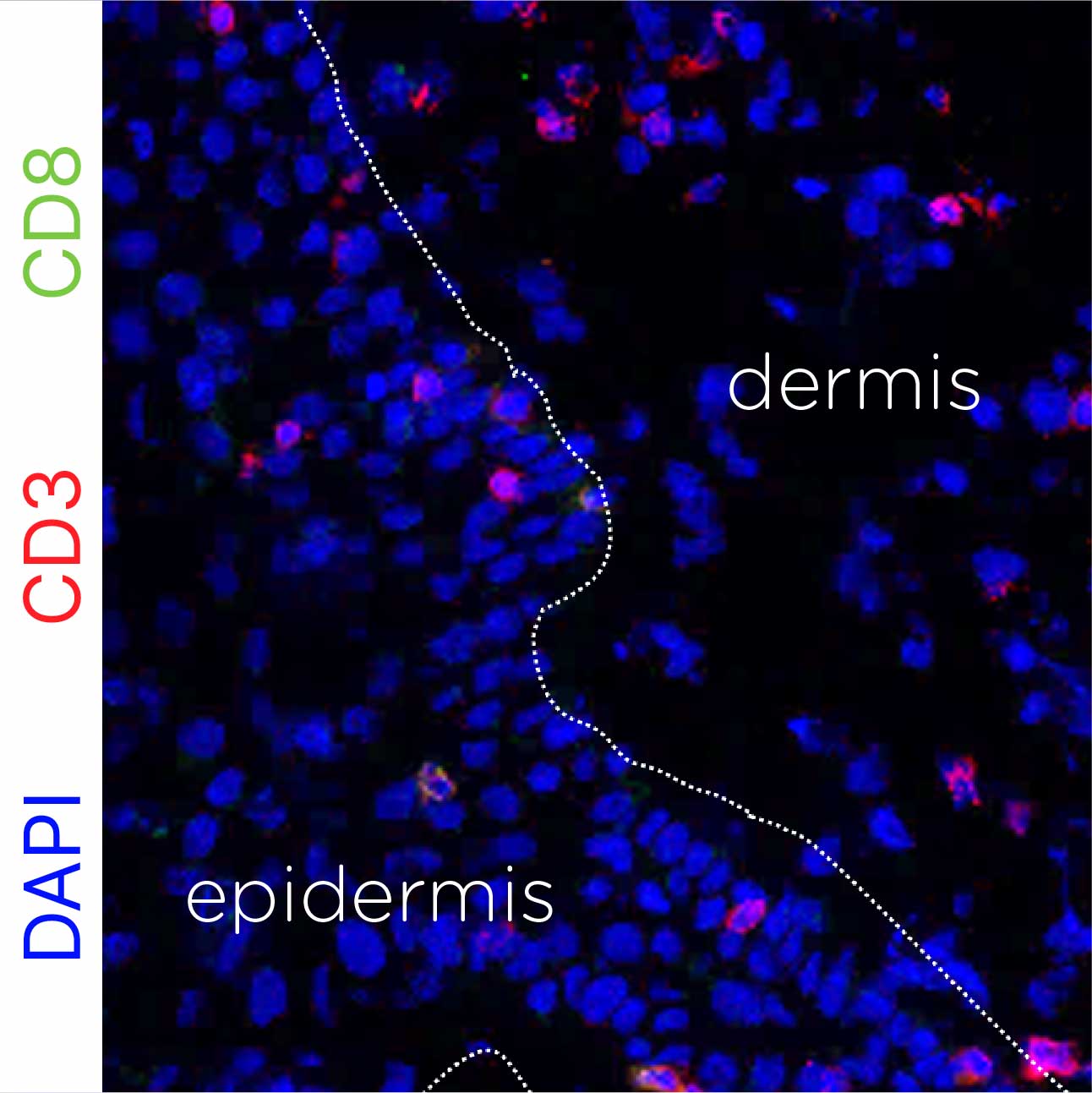
IMMUNE CELL INFILTRATION IN THE DERMIS
Test: Immune cell detection
Method: Immunohistofluorescence staining
Model: Lesional skin biopsies from HS patients
Results: Immunofluorescence staining demonstrates the presence of CD3+ CD8+ T cells infiltrating the dermis of lesional skin from HS patients.
IMMUNE CELL INFILTRATION IN LESIONAL TISSUE FROM HS PATIENTS
Test: Histological Analysis
Method: H&E staining
Model: Lesional skin tissue from patients with HS
Interpretation of results: Representative H&E-stained section illustrating immune cell infiltration (indicated by blue arrows in the top panel). The bottom panel shows a higher-magnification image highlighting nodule formation within lesional tissue.
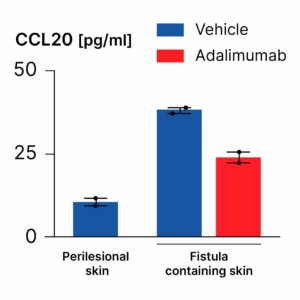
ADALIMUMAB REDUCES CYTOKINE RELEASE IN LESIONAL TISSUE
Test: Cytokine quantification
Method: ELISA test
Model: Organ culture of peri-lesional and fistula-containing lesional skin samples
Interpretation of results: CCL20 levels are significantly increased in the culture medium derived from fistula-containing skin tissue. Treatment with adalimumab markedly decreases the release of CCL20 from skin tissue containing fistulae, indicating an anti-inflammatory effect.


At QIMA Life Sciences, we are committed to staying at the forefront of dermatology research by developing innovative approaches.
We offer smart solutions for studying hidradenitis suppurativa using validated models at both preclinical and clinical stages, making us the perfect partner for your research.
Explore Our Models & Assays in Our Flyer
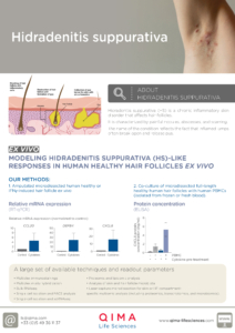
Interested in Learning More?
Explore Other Related Topics
SKIN & HAIR RESEARCH
HIGH-DEFINITION IMAGING: SKINCAM PRO®
