Related Publications
SELECTED PUBLICATIONS ON WOUND HEALING
Breakthrough research identifies new targets for wound healing
Stimulation of the proliferation of human dermal fibroblasts in vitro by a lipidocolloid dressing
Foreskin-isolated keratinocytes provide successful extemporaneous autologous paediatric skin grafts
>> Check the full list HERE.
Conference Contributions
We share our research on dermatology and trichology through international conferences and publications, contributing to the advancement of research in these fields.
>> Check our past contributions HERE.
Preclinical Research Solutions
Clinical Research Solutions
IN VITRO MODELS
- Keratinocytes: NHEK (Normal Human Epidermal Keratinocytes)
- Fibroblasts: NHDF (Normal Human Dermal Fibroblasts), Myofibroblasts, Keloid fibroblast cell line
- Endothelial cells: HUVEC (Human Umbilical Vein Endothelial Cells), HMVEC (Human Microvascular Endothelial Cells)
- RHE (Reconstructed Human Epidermis)
- Dermal equivalent
- Sensory neurons: Primary rat sensory neurons
- Immune cells: Primary mast cells (derived from human skin explants), Polymorphonuclear neutrophils (PMNs), Monocyte-derived macrophages
EX VIVO MODELS
- Human skin ex vivo: “punch-in-a-punch” wound healing model
- Human skin ex vivo: acute & chronic wound healing models
- Human skin ex vivo + S. aureus
- Fresh whole blood
BIOANALYSIS OF CLINICAL SAMPLES
- Non-invasive (swabbing or tape stripping) and invasive (biopsies) sample collection
- Analysis and quantification of cellular components (proteins, lipids) via analytical chemistry
- Wound healing biomarker analysis in tissue, blood and non-invasive collected samples
CLINICAL IMAGING
- Image capture
- Image analysis
CLINICAL TRIALS
- Clinical study implementation
- Clinical study performance
- Data management
- Data analysis
Wound Healing Study Examples
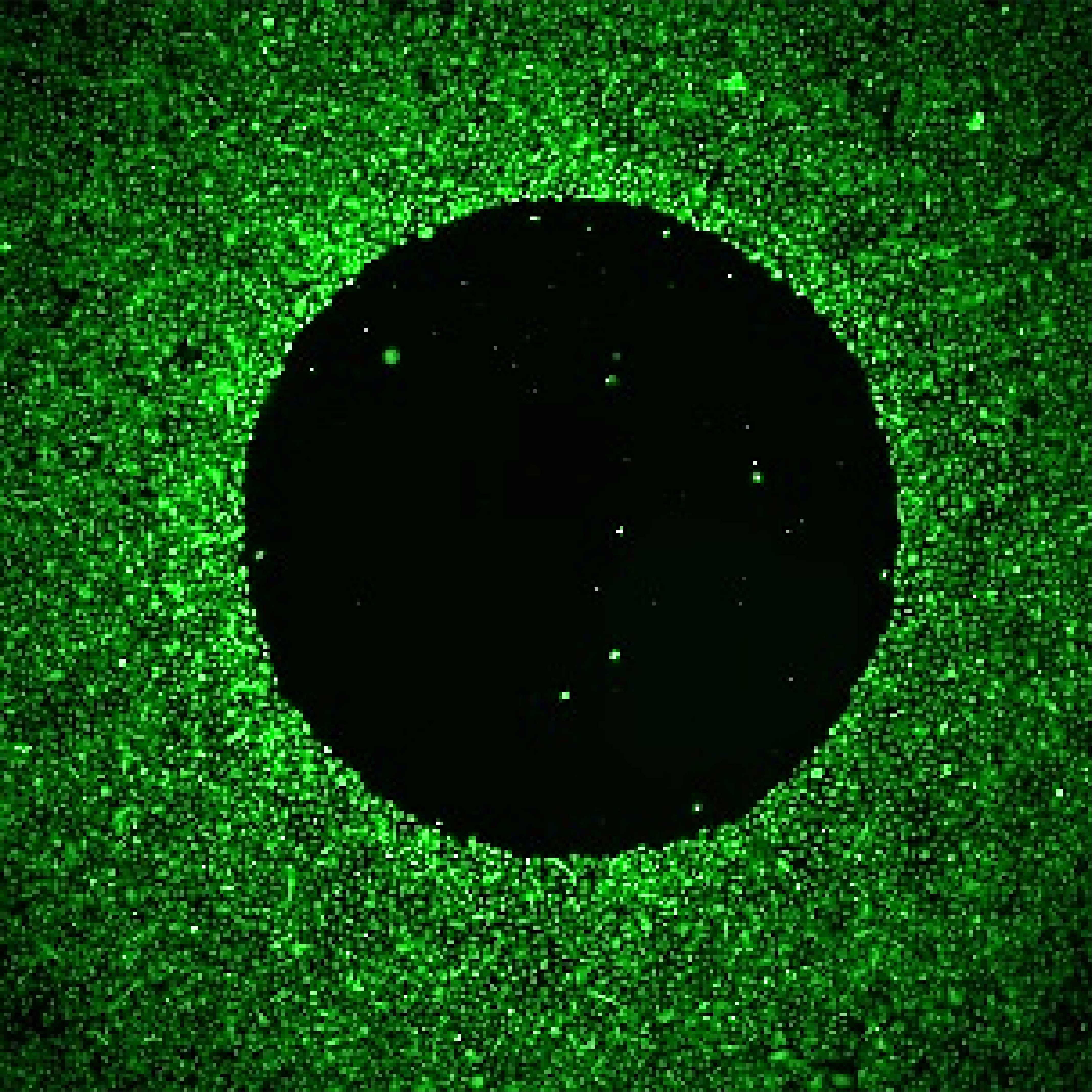
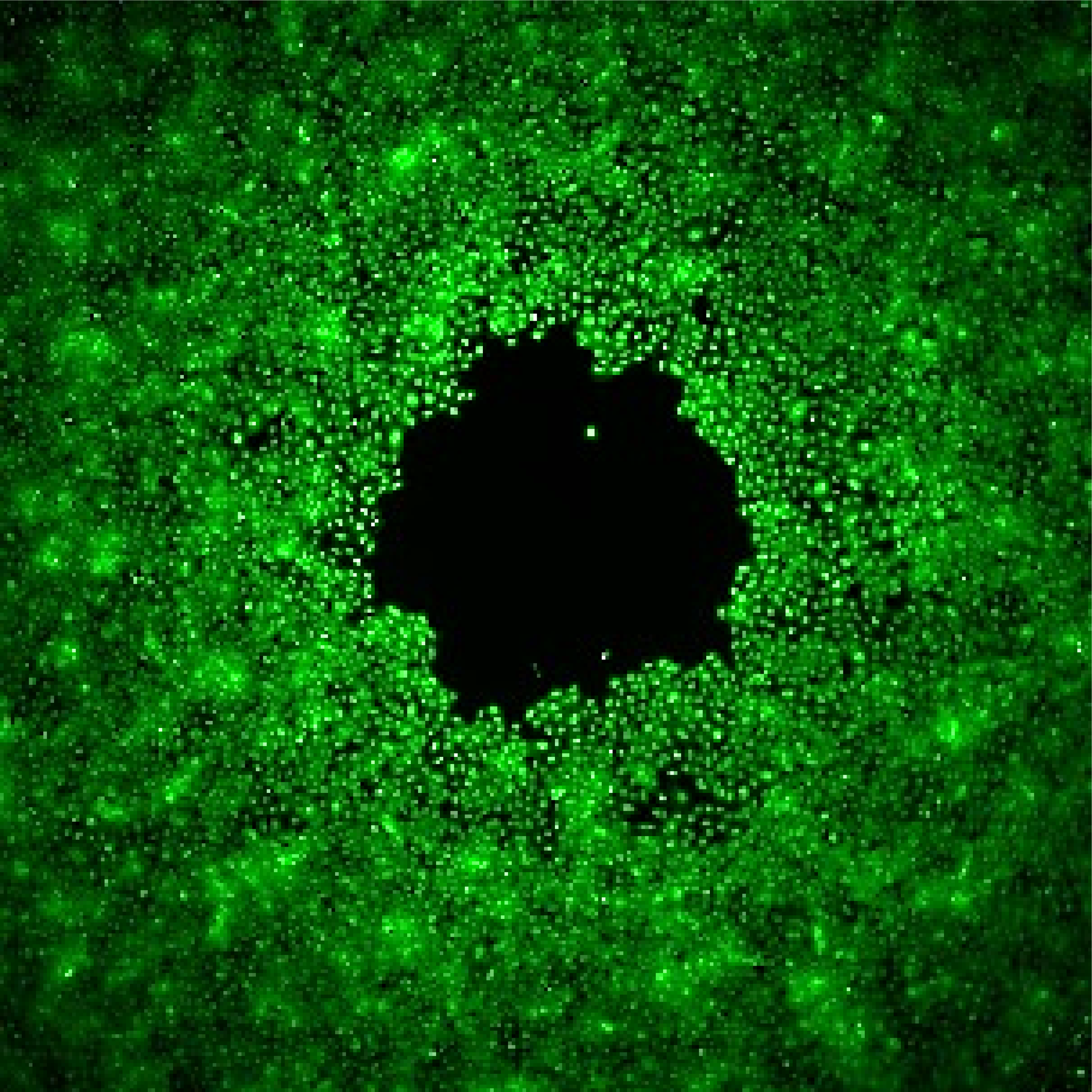
RE-EPITHELIALIZATION
Test: Keratinocyte migration
Interest: This assay quantifies the gap closure rate (the speed at which keratinocytes migrate to cover the cell-free area). This reflects their migratory capacity, a critical factor for successful re-epithelialization.
Method: Fluorescence imaging
Model: Normal Human Epidermal Keratinocytes (NHEK)
Interpretation of results: Over time, keratinocytes migrate to close the cell-free area, significantly reducing the initial ‘wound’ size. Quantitative data not shown.
RE-EPITHELIALIZATION
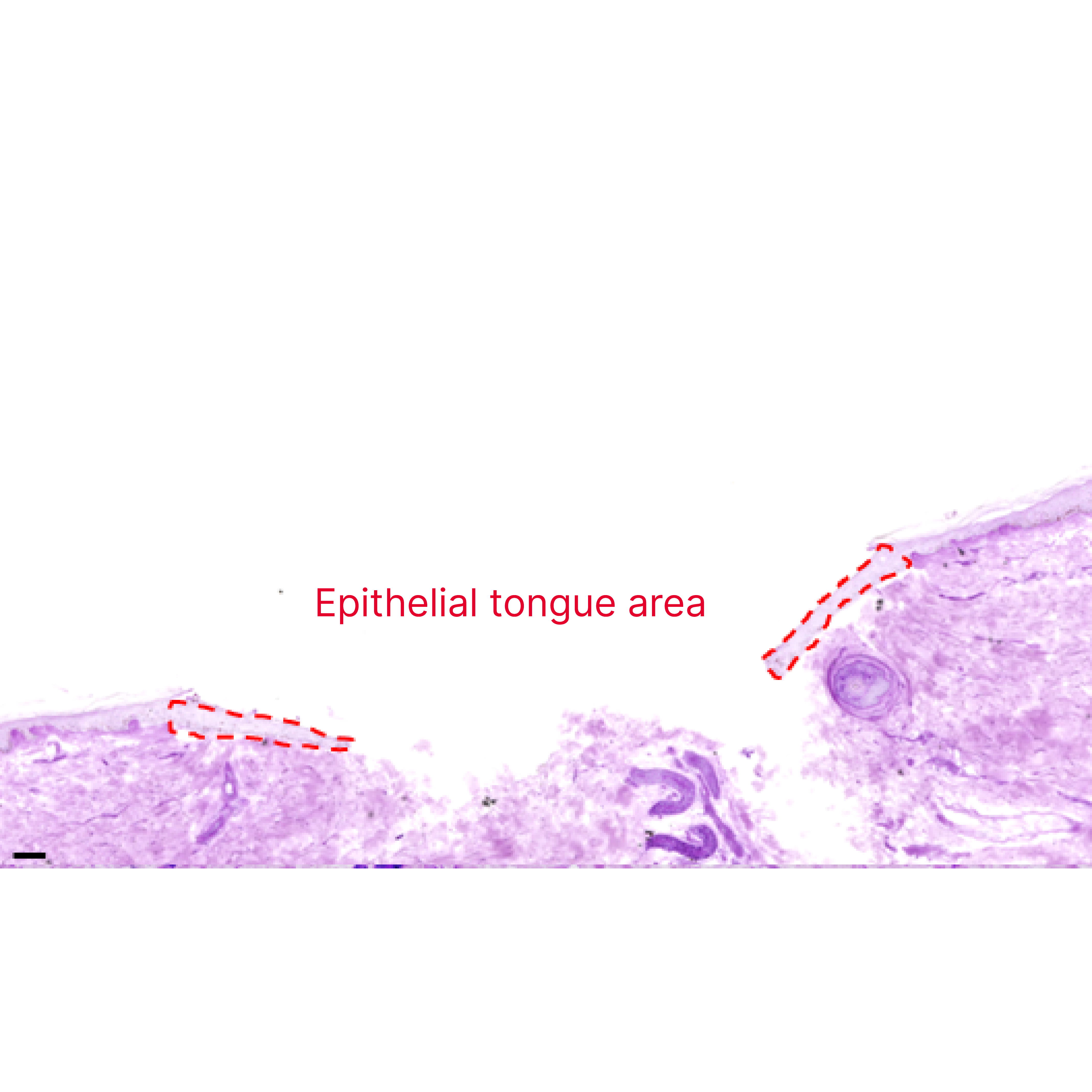
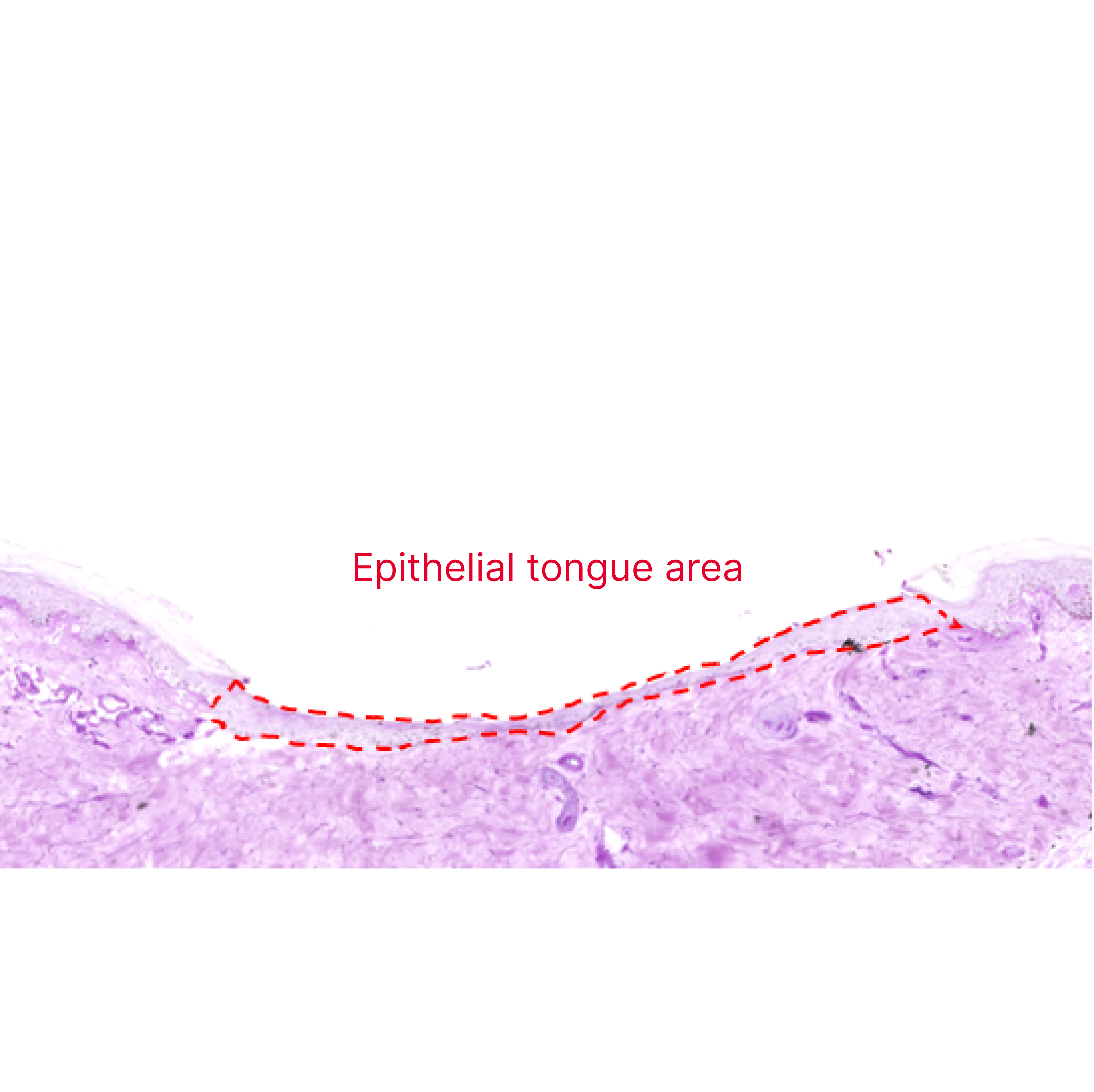
Test: Epithelial tongue length
Interest: The area and length of the growing epithelial tongue provide insights into the migratory and proliferative capacity of keratinocytes, a critical factor for successful re-epithelialization.
Method: Histological image analysis
Model: Human skin ex vivo – “punch-in-a-punch” model
Interpretation of results: Over time, a newly formed epithelial tongue begins migrating to cover the wound bed. Eventually, the entire area becomes covered by a new layer of keratinocytes, completing the process of re-epithelialization.
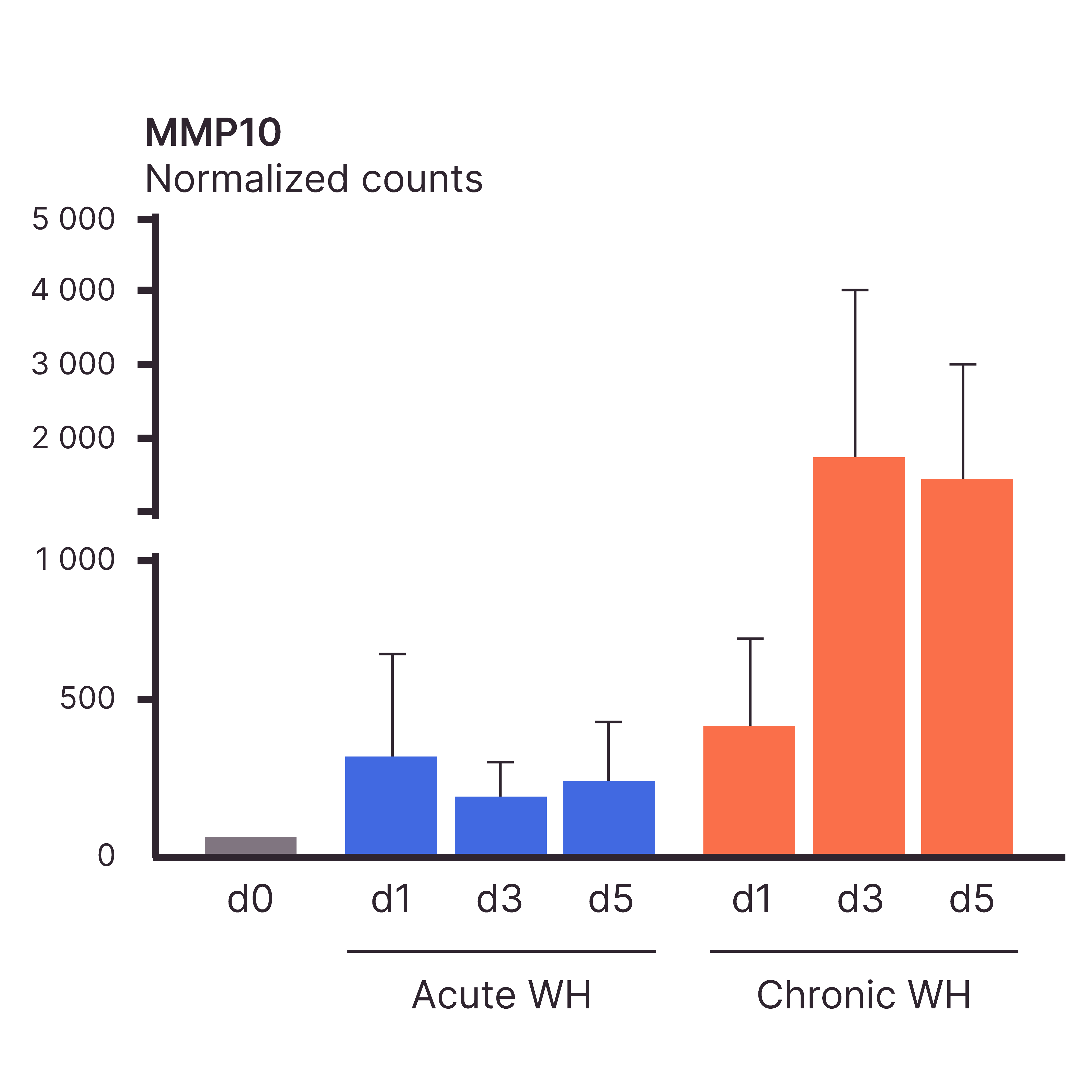
GENE REGULATION IN ACUTE VS. CHRONIC WOUNDS
Test: Wound healing-related gene expression
Interest: Transcriptomic data offers valuable insights into wound healing-related processes and pathways in both acute and chronic wounds. It serves as a vast resource for identifying novel therapeutic targets for wound healing.
Method: Transcriptomics
Model: Human skin ex vivo – acute & chronic wound healing models
Interpretation of results: MMP-10 was strongly upregulated in chronic wounds compared to acute wounds.
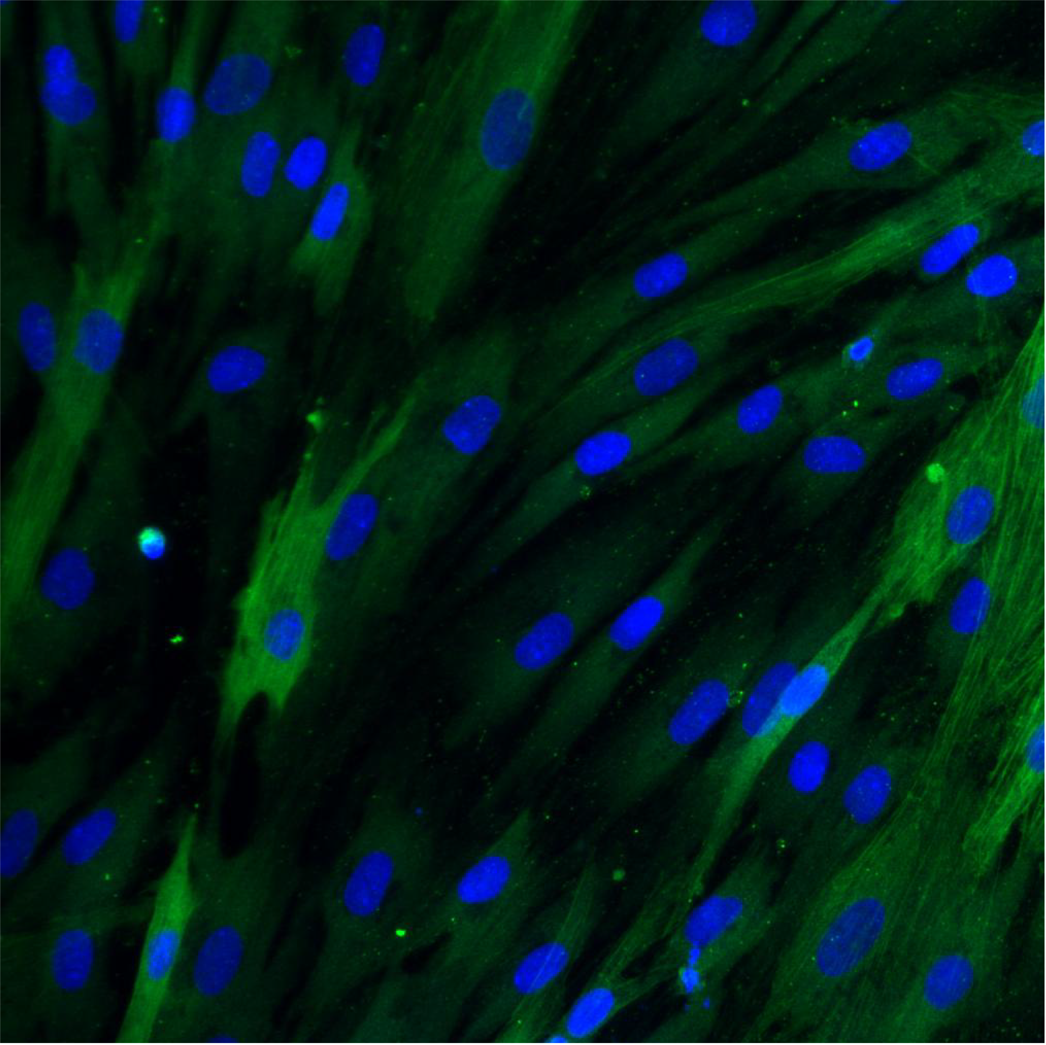
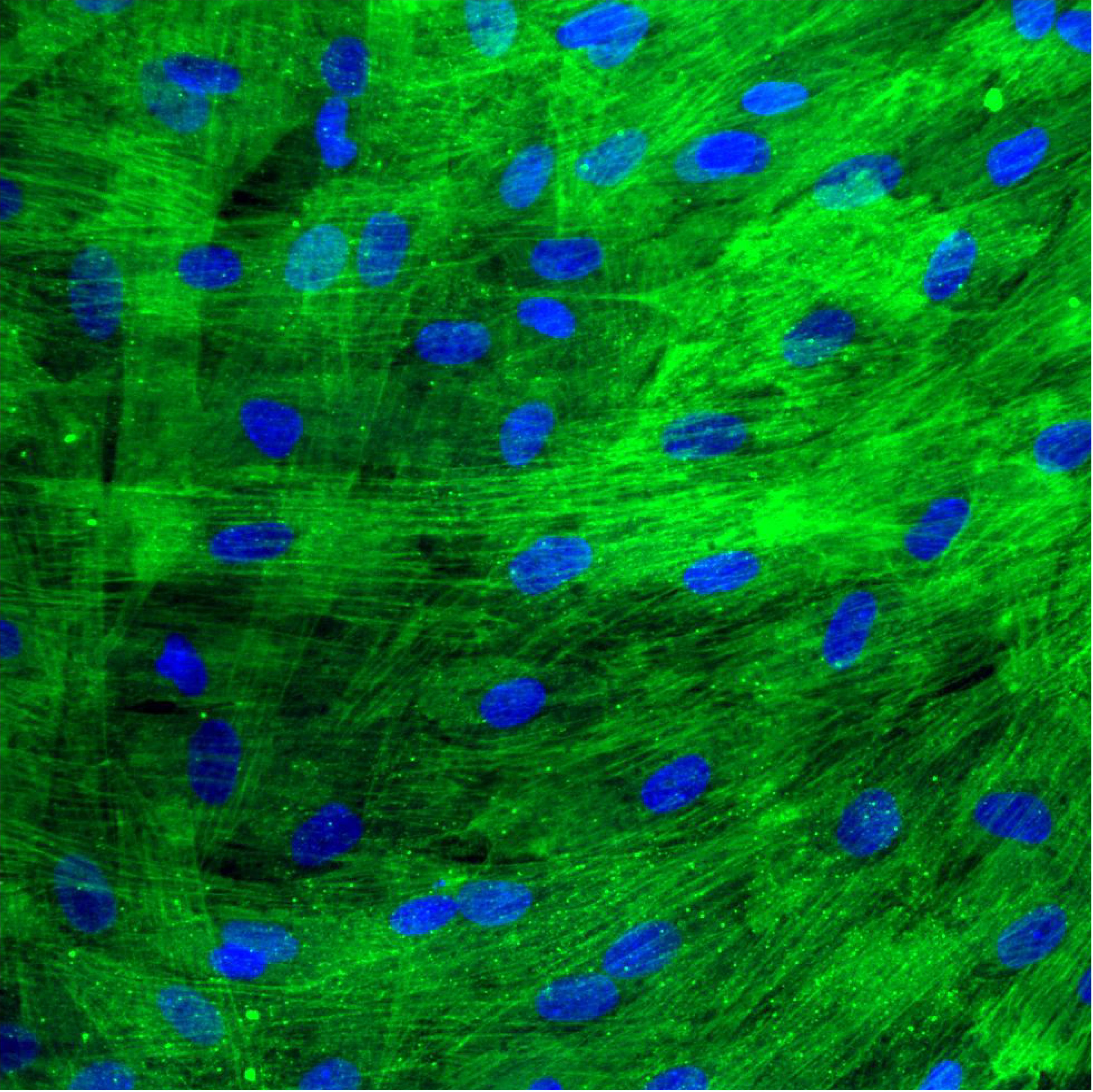
FIBROBLAST DIFFERENTIATION INTO MYOFIBROBLASTS
Test: α-SMA expression
Interest: The differentiation of fibroblasts into myofibroblasts enhances their contractile capacity, allowing them to generate the forces necessary to bring wound edges together and reduce wound size.
Method: Immunofluorescence
Model: Normal Dermal Human Fibroblasts (NHDF)
Interpretation of results: Myofibroblasts exhibit higher α-SMA levels (in green) compared to fibroblasts. Quantitative data not shown.
At QIMA Life Sciences, we are committed to staying at the forefront of dermatology research by developing innovative approaches.
We offer smart solutions for studying wound healing using validated models at both preclinical and clinical stages, making us the perfect partner for your research.
Explore All Our Models & Assays in Our Webinar
Blog Articles
Differences between acute & chronic wounds (upcoming)
Diabetic patients and chronic wounds (upcoming)
What’s New in Testing?
PRECLINICAL SOLUTIONS
Primary mast cells (derived from human skin explants)
Chronic & Acute wound healing ex vivo models
Transcriptomic profiling of chronic and acute wounds
Cell line with keloid fibroblast-like morphology


