


QIMA Life Sciences has several canine in vitro models which were initially developed in cooperation with veterinary practitioners. These models are 3Rs aligned, and have been validated for modelling various pathologies and stressors, and for testing the tolerance and safety of veterinary treatments and pet care products alike.
Our reconstructed Canine Epidermis (RCE) is a 3D epithelial model developed from primary canine keratinocytes. It is designed to recapitulate key structural and functional features of native canine epidermis. For atopic dermatitis (AD) applications, a characterised AD-like state (RCE-AD) can be reproducibly induced in the model, using a defined cytokine cocktail.
The data below illustrate model responsiveness across four readout categories, using a reference molecule as a positive control. Please do not hesitate to contact us for more information.
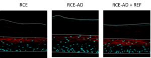
1 – Histomorphology: cytokine induction drives spongiosis and architectural disruption.
Analysis of the morphology by histological staining highlights a pro-inflammatory effect of the cytokine cocktail (increased spongiosis, disruption of the basal layer architecture, and a decrease of keratohyaline granules), the reference compound counteracts this effect.
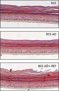
Histological morphological analysis hemalun eosin staining
2 – Barrier permeability: cytokine induction increases epidermal permeability, inhibited by reference compound.
The analysis of the permeability of the membrane can carried out by the quantification of ceramides by LC/MS and by the evaluation of the penetration of the Lucifer yellow within the corneous layer, among other methods. Barrier integrity may also be assessed by electrical resistance and transepidermal water loss.
The quantification of ceramides by LC/MS shows a reduction in ceramide quantities in the RCE-AD model; in this case the reference compound does not mitigate the pro-inflammatory effect of the cytokine cocktail.
The evaluation of the penetration of Lucifer yellow shows that the presence of cytokines (RCE-AD) increases the permeability of the tissue and promotes the penetration of Lucifer yellow. The reference inhibits the effect of cytokines on the epidermal barrier permeability.
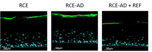
Evaluation of the penetration of Lucifer yellow into the RCE
See more on the RCE model

3 – Epidermal proteins: filaggrin, corneodesmosin and claudin-1 are downregulated by cytokine induction.
Skin barrier function is also frequently evaluated in our RCE models by analyzing the expression of epidermal and tight junction proteins. Differentiation proteins (Filaggrin) and adhesion proteins (Corneodesmosin) are analyzed by WES (Automated Western Blot) and/or immunolabelling. Tight junctions are quantified by immunolabeling. RT-qPCR may also be used to profile tissue responses to stimuli.
The evaluation of epidermal proteins and tight junctions shows that the cytokine cocktail (RCE-AD) strongly decreases the expression of profilaggrin, Corneodesmosine and Claudin-1. The reference significantly decreases the effect of cytokines on the expression of these proteins.
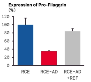
Profilaggrin expression
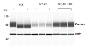
Evaluation of Corneodesmosin expression – WES
Evaluation of Claudin-1 expression by immunolabeling
4 – Inflammatory readout: IL-8 secretion is robustly elevated in RCE-AD and reduced by reference compound.
Cytokine profiling in the RCE and RCE-AD models are useful tools for evaluating inflammation, skin irritation, and disease-like states. Multiple endpoints have been characterised in these models.
For example, when inflammation is evaluated by ELISA quantification of IL-8 in the medium, there is a strong increase of IL-8 secretion in RCE-AD, and a decrease of this response with the reference treatment.
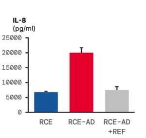
Evaluation of inflammation by ELISA quantification of IL-8
Summary
The RCE-AD model delivers a mechanistically coherent, multi-endpoint readout profile across barrier integrity, epidermal protein expression and inflammatory mediator secretion; providing three dimensional tissue-level resolution. The data shown here represent one configuration; the model is adaptable to a range of therapeutic hypotheses, candidate classes and custom readout panels.
Beyond atopic dermatitis, the reconstructed canine epidermis platform supports topical safety and tolerance profiling, modelling defined dermatological challenges (inflammation, infection, stressors etc.), and co-culture studies — making it a versatile substrate across veterinary dermatology research programmes.
For more model capabilities, example data, and platform applications, download our RCE flyer or contact our scientists directly to discuss fit for your programme.
See also


