


Share this Technical Focus
Key takeaway:
Our senescent melanocyte (SenMel) model enables the study of cellular aging processes in melanocytes.
Also, it offers valuable insights into the mechanisms behind, providing a platform for assessing the impact of cosmetic actives on skin aging and pigmentation.
Melanocytes are pigment-producing cells that determine skin color and play a major part in protecting the skin against ultraviolet radiation.
Existing research rarely explores how baseline pigmentation levels may influence melanocyte susceptibility or adaptive responses to oxidative stress, one of the main factors contributing to cellular senescence.
To address this lack of data, we developed the SenMel model, which induces melanocyte senescence in vitro through chronic exposure to hydrogen peroxide (H₂O₂).
Access to our
Human Senescent Melanocytes Poster

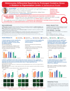
Evaluation of Cellular Senescence in Melanocytes
Test: Melanocyte Senescence
Method: Senescence Associated Beta-galactosidase Activity
Observation: Chronic exposure to H₂O₂ promotes a senescent phenotype in all melanocytes, regardless of their baseline pigmentation, demonstrating the broad applicability of the model.
Melanocyte Morphological Alterations
Test: Cell Morphology
Method: Immunofluorescence staining
Observation: Aged melanocytes treated with IBMX, and co-cultured with keratinocytes, show fewer dendrites and appear flatter after chronic H₂O₂ exposure. In comparison to the control condition, these melanocytes exhibit more pronounced features of aged cells.
Increased Sensitivity to the Induction of Melanosome Transfer
Test: Melanosome Transfer
Method: Flow cytometry
Observation: Under control conditions, both young and senescent (H₂O₂) groups exhibited higher melanosome levels within melanocytes compared to keratinocytes. After stimulation, both young and H₂O₂-induced senescent groups showed an increased percentage of melanosomes in keratinocytes, surpassing those in melanocytes.
*Pmel17 is a melanosome-specific structural protein. Percentage of positive cells were calculated out of the total number and keratinocytes and melanocytes present in the sample.


Written by:
Emmy Vanotti
Scientific Communication & Marketing Project Leader
