


About Melanoma
Melanoma is a skin tumor caused by the uncontrolled proliferation of melanocytes. Although its incidence is relatively low—affecting only 3 to 5 individuals per 100,000—it accounts for 75% of skin cancer-related deaths due to its rapid progression and aggressive spread. This malignancy often arises from physical or chemical damage to the skin, such as intense sun exposure. In most cases, melanoma is detected when a dark-colored spot appears on the skin (80% of cases) or when a pre-existing mole changes in color or shape.
Melanomas progress through three distinct stages:
- Radial Growth Phase (RGP): Melanocytes proliferate abnormally in a horizontal pattern, remaining along the basal lamina.
- Vertical Growth Phase (VGP): Cell proliferation accelerates, enabling vertical expansion into the dermis. This is driven by the significant release of metalloproteinases, proteolytic enzymes that degrade the extracellular matrix and basal membrane, facilitating melanoma invasion.
- Metastatic Stage: In this final phase, melanoma cells acquire the ability to intravasate, allowing them to spread through the bloodstream or lymphatic system, forming metastases in other organs.
The mechanisms governing the transition between these stages remain poorly understood. However, research has identified several genes involved in these critical shifts
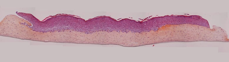
Figure 1: Reconstructed skin model with melanoma
In vitro Models of Melanoma
QIMA Life Sciences has launched a tissue engineering R&D program to develop a skin-equivalent model incorporating melanoma cells.
After the testing of various monolayer cultures, two melanoma-derived cancer cell lines from QIMA Life Sciences’ collection were selected for integration as standard components in a normal skin-equivalent model (3D culture):
- A-375 (ATCC® CRL-1619): Cells derived from metastatic melanoma
- WM266-4 (ATCC® CRL-1676): Cells derived from metastatic melanoma
3D cell culture has allowed us to obtain 2 models corresponding to different stages of development and severity, in which:
- The A-375 line presents an aggressive profile and results in very important epidermis destruction and equivalent dermis invasion over culture time
- The WM266-4 line develops a less aggressive response: after 10 to 12 days of culture, the epidermis part of reconstructed skin shows fairly good histology and cancer cells form clusters that are easily identifiable in histology. The cluster size increases over culture time.
Prospects
Our objective is to provide customized, coherent models and methods to address the challenges faced by research and industry projects related to melanoma. We now offer the opportunity to use various models directly, either for pharmacological studies or as part of collaborative R&D programs in melanoma research.
We currently offer analytical solutions in cellular and molecular pharmacology to facilitate rapid project completion while also advancing the development of new analytical methods.
