Over the last decades, scientists have developed and continued to improve 3D cell culture techniques, leading to the emergence of new models.
These cutting-edge models have revolutionized the compound testing paradigm.
Now, compounds can be assessed in a 3D cellular context, which more closely mimics the physiological organization of cells within the human body.
What Are 3D In Vitro Models?
Most frequently cells are cultivated on a flat surface as a monolayer. This method is called 3D culture.
But if cultured under specific conditions, cells can organize in a different way. They can grow into a three dimensional structure. At this point, cells no longer interact with the extracellular matrix in just two dimensions, but in three dimensions, which more closely resembles their natural organization within organs.
These 3D in vitro models can be developed using various methods, either by culturing cells
- on a scaffold (such as hydrogel or inert matrix), or
- scaffold-free, using specific culture vessels.
Also, the cells used in these models can be derived from various sources, including:
- human or animal tissues, or
- cell lines, primary cells, stem cells, or patient-derived cells.
Learn more about in vitro models
Our 3D In Vitro Models
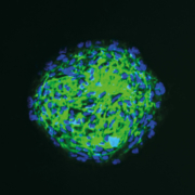
Hair spheroids & organoids
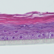
Reconstructed Human Epidermis (RHE)
Dermal Equivalent (DE)
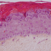
Reconstructed full-thickness human skin
The donor’s genetic background and health status can influence the characteristics of primary cells, while culture conditions can affect the characteristics both primary cells and cell lines.
Our models can be thus categorized as:
- “healthy”,
- “pathological”, and
- “mimicking pathological conditions”.
Our Extensive Array Of Assays
We provide a wide range of assays on various 2D in vitro models.
Here are some examples of readouts we can assess:
- Cytotoxicity
- Oxidative stress markers
- Inflammation markers
- Cell proliferation
- Signal transduction
- DNA damage
- Cell migration
- Cell differentiation
- Melanogenesis
- Lipidogenesis
- …among many others!
Why Use 3D In Vitro Models?
The central advantage of 3D cell culture lies exactly where 2D models fail: the ability to accurately represent the architectural organization of human tissue outside the body.
3D cell culture systems are more physiologically relevant, offer better prediсtivity, and retain a “steady state” for longer, due to their greater structural complexity.
In addition, 3D cell culture offers an opportunity to:
- Study interaction between different types of cells
- Integrate the flow of biological fluids
- Represent barrier tissues, such as the epithelium
- Simulate the growth of cells and organs
- Grow and treat tumor cells, including cancer
- Useful for testing finished products (e.g., topical application of a cream onto the surface of a reconstructed epidermis)
You want to learn more about our 3D in vitro models?
Click on the button below and let’s have a chat!


