
Atopic dermatitis – initiation and activation step
Initiation: keratinocyte activation and TSLP release
The skin lesions facilitate the passage of an antigen that the subject has previously been sensitized to. This antigen may be mimicked by a microbial pattern or a double stranded viral RNA, particularly poly(I:C) which can be recognized by receptors such as the Toll-Like Receptor 3 (TLR3). Moreover, the same lesions cause the membrane of certain keratinocytes to rupture, leading to a release of cytoplasmic IL-1α that may bond with the IL-1R receptors located on the neighboring keratinocytes. This double stimulation by an antigen (poly(I:C)) and cytokines (IL-1α) causes specific keratinocyte signaling pathways (NFκB, MAP kinases, etc.) to become activated, leading to early expression of TSLP (key cytokine of AD [Test : EPIBA-0070]), and then of chemokines that enable the recruitment of the various cells involved in AD [Tests : NHEK-0090; EPIBA-0069]. TSLP subsequently activates various cell populations (mast cells, plasma cells, basophil cells and macrophage cells) and predominantly, dendritic cells (DC) via the TSLP-R/IL-7Rα heterodimeric receptor.
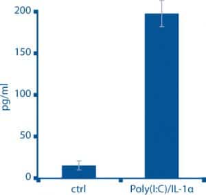
NHEK, TSLP release (stimulation of poly (I:C) + IL-1 alpha)
[Assay : NHEK-0091]
Dendritic cell activation and T Lymphocyte polarization
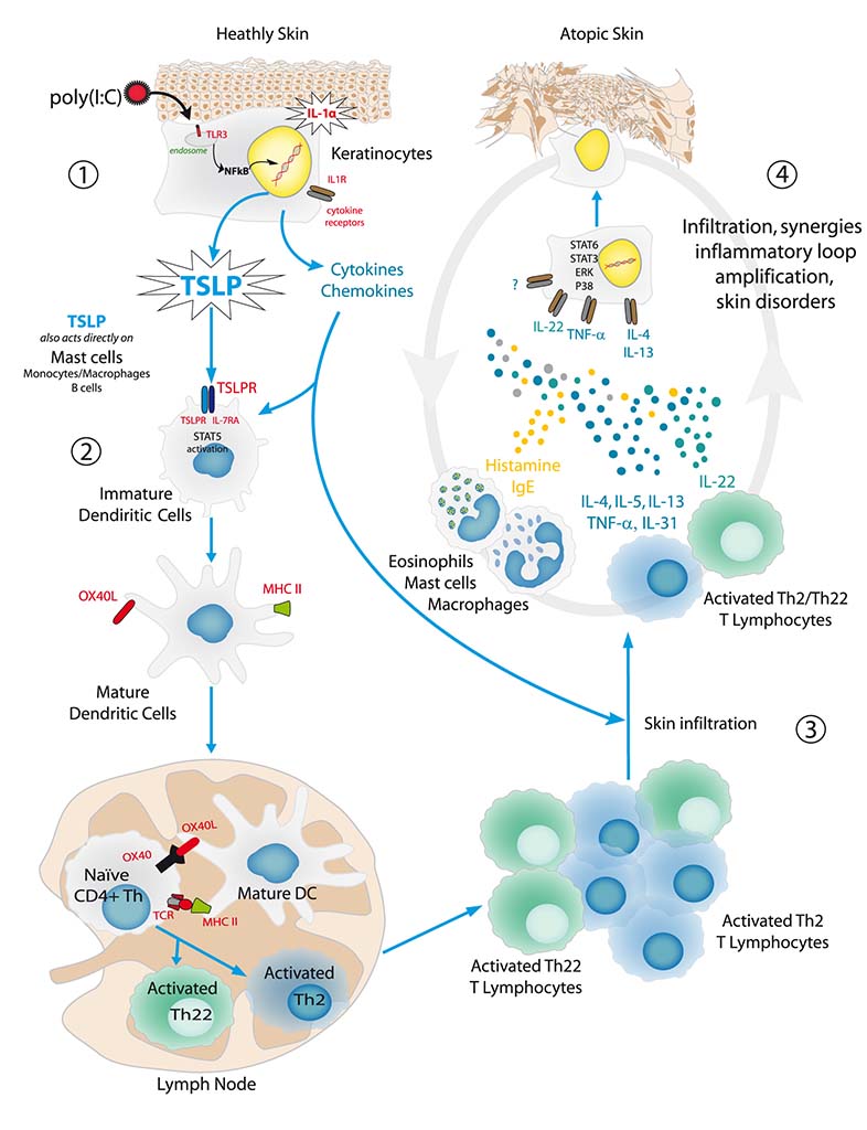
After activation by TSLP, DC proliferate and differentiate into mature DC capable of producing numerous chemokines and innate immunity molecules. These mature cells express type II major histocompatibility complex (MHC II) and OX40L ligand on their surfaces. DC then migrate to the lymph nodes where the antigen is presented to the immature T lymphocytes (CD4+ TL). This stage is carried out by an interaction between MHC II and the T Cell Receptor (TCR) of the TL, and by the binding of OX40L with its receptor located on the surface of the TL. The presentation of the antigen enables the polarization of CD4+ TL into TL of type Th2 and Th22, which are essential to the immune response of AD [Test : CD4TL-0005]. This Th2/Th22 dominant immune response is characteristic of atopic dermatitis.
References
- Cho, SH., et al. (2001). J Invest Dermatol. 116:269-274.
- Odhiambo, JA., et al. (2009). J Allergy Clin Immunol. 124(6):1251-1258.
- Oyoshi, MK., et al. (2009). Adv Immunol. 102:135-226.
- Spergel, JM., et al. (2010). Ann Allergy Asthma Immunol. 105:99-106.
- Schlievert, PM., et al. (2010). J Allergy Clin Immunol 125:39-49.
- Boguniewicz, M., et al. (2010). J Allergy Clin Immunol. 125:4-13.
- Palmer, CN., et al. (2006). Nat Genet. 38:441-6.
- Kinoshita, H. et al. (2009). J Allergy Clin Immunol. 123:179-86.
- Anh, T. et al. (2011). J Invest Dermatol. 131:2205-2212.
Related posts


Check out Bioalternatives’ updates and experience new testing ideas
- New assays, models and services
- Posts and publications
- Events

