in vitro models & assays
QIMA Life Sciences has many in vitro or ex vivo models at your disposal:
- melanocyte cell lines (B16, etc.)
- normal human epidermal melanocytes (NHEM)
- co-culture of normal human epidermal keratinocytes and melanocytes (NHEK/NHEM)
- melanocyte-containing reconstructed epidermis (RHEm)
- skin explants (ex vivo)
- hair follicle (ex vivo)
on which we can evaluate the whitening or pro-pigmenting effect of active cosmetic compounds or formulations by measuring:
- the expression of specific markers of melanogenesis: TYR, TRP-1, Pmel17, MITF, etc.
-
Expression and phosphorylation of the master transcription factor regulating pigmentation
- tyrosinase activity and expression
- melanin synthesis
- melanocyte dendricity
- Melanocyte differentiation
- melanosome transfer
- Number of mature and immature melanocytes
- Expression of growth factors regulating pigmentation and corresponding receptors
Data mapping and clinical imaging
Measurement of skin pigment imperfections in 2D color imaging
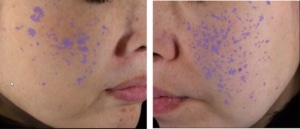
Signs of photo-ageing, such as solar lentigo, can be detected and analyzed by image analysis, either on the whole face or on the back of the hand, or by targeting a specific lesion.
According to the area to be investigated, a choice will be made regarding the type of shot for the whole face, the hand, or a small sun-exposed area of the body (e.g., the upper chest).
Other pigment lesions, such as melasma and post-inflammatory pigmentation are also analyzed in the same conditions.
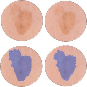
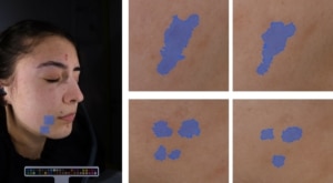
The density of pigmentation spots, their surface, their color intensity, and the contrast measured with surrounding skin, as well as homogeneity of complexion are all parameters that can be extracted from this analysis.

ColorFace®


Analysis of solar lentigos on the whole face, with images acquired with ColorFace®

Analysis of the PIH of whole face on images acquired with ColorFace ®

DigiCam

Image acquisition of back of hand with DigiCam® and analysis of pigmentation spots

SkinCam
 Image acquisition of temple with SkinCam®
Measurement of melanin in hyperspectral imaging
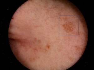
Investigation of sun-induced pigmentation, which is non-invisible but will appear with age, can be performed by using UV polarized monochrome imaging. Parameters such as lesion surface and density can be extracted and monitored over time.
Measurement of melanin in hyperspectral imaging
Newtone’s hyperspectral imaging is a unique technology developed to calculate the skin chromophore absorption mappings, and consequently to measure evolution over time, while providing a perfectly representative image of the measured evolution.
Acquisition systems allow image acquisition of the whole face (SpectraFace) or of a defined area of the body and face (SpectraCam).
On these images, pigment lesions can be monitored and analyzed as by color imaging, while discriminating melanin and hemoglobin.


Hyperspectral imaging


