
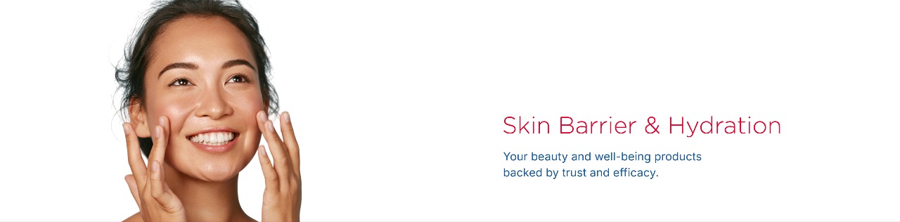
The skin barrier is a multifunctional structure essential for maintaining homeostasis. It limits transepidermal water loss (TEWL), shields the body from external aggressors, and modulates immune responses. Its integrity depends on a complex interplay between corneocytes, intercellular lipids (notably ceramides, cholesterol, and free fatty acids), tight junctions, (corneo)desmosomes, and the underlying epidermal renewal processes.
Hydration is a key component of barrier function. Natural Moisturizing Factors (NMFs), aquaporins, and an organized lipid matrix help the skin retain moisture and maintain suppleness. When disrupted – by cold, UV, surfactants, pollutants, or aging– these systems lose efficiency, leading to dehydration, discomfort, sensitivity, and inflammation.
A healthy barrier is also a resilient barrier. Its ability to recover after stress depends on the coordinated action of keratinocyte proliferation, differentiation, and lipid synthesis. Supporting this regeneration process is critical to improving skin comfort and preventing premature aging.
At QIMA Life Sciences, we provide advanced tools to assess hydration levels, barrier function, and recovery dynamics.
We Support You at Every Stage of the Innovation Cycle to Substantiate Efficacy Claims
Our Skin Barrier & Hydration Preclinical Solutions

IN VITRO MODELS
- NHEK (Normal Human Epidermal Keratinocytes)
- NHDF (Normal Human Dermal Fibroblasts)
- Sebocyte Cell Line SEBO662
- Sebocyte Cell Line SEBO662AR (androgen-sensitive)
- RHE (Reconstructed Human Epidermis)
- RHE displaying barrier defects
- Reconstructed Full-Thickness Human Skin
- 3D Sebocyte Model

EX VIVO MODELS
- Human Skin Explants (Ex vivo skin)
Barrier Default Models:
PRIMARILY BASED ON:
- Pharmacological induction: A cytokine cocktail can be applied to our models to simulate inflammation-associated barrier dysfunction.
- Genetic engineering: Filaggrin deficiency can be induced via gene silencing (siRNA).
- Physical stress: Barrier dehydration can be modeled by culturing cells or tissues in a low-humidity (dry) environment.
We can help you substantiate skin barrier and hydration claims
Reduced TEWL
Promotion of Epidermal Differentiation
Increase in Filaggrin Expression
Increase in Long-chain Ceramides Levels
Enhanced expression of key skin barrier proteins (involucrin, SPRPs, loricrin)
Improved cell junction integrity
Epidermal lipid profiling
Increase of ECM-derived hydration factors (NMFs)
Our Skin Barrier & Hydration Clinical Solutions

BIOANALYSIS OF CLINICAL SAMPLES
- Sample collection (swabbing or tape stripping)
- Analysis and quantification of cellular components (epidermal lipids, including ceramides, cholesterol and free fatty acids) via analytical chemistry
- Analysis of differentiation markers & other biomarkers
- Skin surface analysis via electron microscopy

CLINICAL IMAGING
- Image capture
- Image analysis (e.g., skin roughness)
- Illustration services (e.g., hydration mapping)

CLINICAL TRIALS
- Clinical study implementation
- Clinical study performance
- Data management
- Data analysis
We can help you substantiate skin aging-related claims
Skin Barrier Repair
Skin Soothing
Redness & Inflammation Reduction
Moisture Loss Prevention
Deep Hydration & Nourishment
Reinforces the skin’s natural barrier
Gentle on sensitive skin
Skin Barrier & Hydration Study Examples
PROTEIN STRUCTURE
Test: Filaggrin expression
Interest: Filaggrin and involucrin are essential for the structural integrity and hydration of the skin barrier, providing protection against environmental threats.
Method: WES
Models: RHE and RHE sh-FLG (RHE with barrier default)
Interpretation of results: We successfully developed a RHE model with an impaired skin barrier, as characterized (among other parameters) by a significant decrease in involucrin and filaggrin levels.
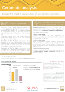
LIPID STRUCTURE & CEMENT
Test: α-hydroxylated ceramides analysis
Interest: Ceramides are key lipids in the skin barrier because they maintain its integrity, hydration, and ensure its protection against environmental stressors.
Method: Liquid chromatography–mass spectrometry (LC–MS)
Model: RHE
Interpretation of results: Vitamin C promotes the synthesis of α-hydroxylated ceramides, enhancing skin barrier function, whereas retinoic acid reduces their levels, potentially compromising the barrier’s integrity.
EPIDERMAL STRUCTURE
Test: Epidermal structure
Interest: Hydration impacts the epidermal structure and overall skin architecture
Method: Hematoxylin-eosin staining
Model: RHE cultured under different humidity conditions
Interpretation of results: RHE cultured in a dry atmosphere exhibited reduced epidermal thickness and a decreased number of keratohyalin granules.
CLINICAL SIGNS OF HYDRATION
Test: Skin microrelief (roughness)
Interest: Dehydrated skin exhibits a rough texture, often characterized by fine scaling, flakiness, and accentuated microrelief.
Method: Image capture (SkinCam®) + Microrelief analysis
Model: Human volunteers
Interpretation of results: After 7 days, the treatment demonstrated clinical efficacy, evidenced by a reduction in skin roughness and dryness.


At QIMA Life Sciences, we specialize in providing comprehensive solutions for studying skin barrier and hydration through validated models at both preclinical and clinical levels.
Our dedication to scientific innovation drives us to develop cutting-edge models and approaches, staying aligned with the latest advancements in skin research.
Explore All Our Models & Tests in Our Brochure

Related Publications
What’s New in Testing?
OUR SOLUTIONS
Skin-Biosense® to monitor skin barrier integrity
Analysis of epidermal ceramides
Blog Articles
Interested in Learning More?
Explore Other Related Topics
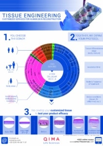
TISSUE ENGINEERING
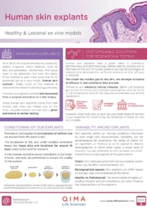
HUMAN SKIN EXPLANTS
FACE IMAGING SYSTEM: SKINCAM PRO®
CERAMIDES ANALYSIS
SKIN BARRIER & HYDRATION
Updated June 1, 2026
