
Hair: Follicle, Associated Structures and Growth
A person’s hair is often considered as a remnant of the animal mane and continues to have both a social role (as a symbol of youth, health and fertility) and a biological role (by protecting the top of the head from the sun and by the sequestration and excretion of unwanted biochemical substances). Hair is a unique feature of each individual and, more generally, of the human species. It is estimated that adults have about 5 million hair follicles, with 1 million hair follicles found on the head, and with only 120,000 to 150,000 hair follicles covering the scalp. Depending on the region of the scalp, their density varies by 200 to 300 follicles /cm2. A hundred hair follicles are naturally shed each day. The speed at which hair grows is about 0.3 mm per day or 12 cm per year. This speed is affected by numerous factors, such as follicle location, gender, age and ethnicity of the individual as well as environmental factors.
Follicle and associated structures
Hair follicles are skin adnexa resulting from epithelial-mesenchymal interactions programmed in the third month of embryonic development. These miniature organs are able to regenerate in a complete, autonomous, cyclic and asynchronous manner that is unique in the human body. They regenerate more than 8 to 10 times over a lifetime. The hair follicle root, which is located 4mm under the epidermis, includes sebaceous glands and interacts with the associated structures (epidermis, dermis, arrector pili muscles, vascular system). The histological analysis of the pilosebaceous unit reveals a three-element organisation: the epithelial compartment, the mesenchymal compartment and the sebaceous gland.
The epithelial compartment
It is composed of the matrix, hair shaft, inner sheath and outer epithelial sheath.
Matrix
The matrix, which is located at the base of the follicle and surrounds the dermal papilla, contains highly proliferative and only slightly differentiated cells. These cells are the source of the different cellular types making up the follicle [Assays: FOLL-0003; FOLL-0004].
Hair shaft
This is the visible structure of the hair and is composed of three concentric layers (the cuticle, cortex and medulla).
- The cuticle, the 3.5 to 4.5 µm outer layer, is composed of cells held together by a ceramide-rich cement, which provides protection for the basal layers.
- The cortex, which is the middle layer and represents 75% of the total diameter, is the main body of the hair. This region is primarily composed of keratin, which is responsible for hair cohesion, stiffness and suppleness. It contains the melanin pigments that give hair its colour [Assays: FOLL-0011; FOLL-0015]. This production depends on the expression and regulation of key genes [Assay: FOLL-0009].
- The medulla, or medullary canal, is the central part. The hair shaft, which is partially controlled by transcription factors LεF-1/CTF, very specifically expresses transglutaminase-3 as well as 15 keratins [5]. The expression levels of these markers can be evaluated by transcriptome analysis [Assay: FOLL-0004] or immunohistological analysis [Assay: FOLL-0003].
Inner and outer epithelial sheathes
The inner sheath, which is composed of three concentric layers (referred to as Henle’s layer, Huxley’s layer and cuticle), is keratinised. The differentiation of its cells partially depends on the GATA-3 transcription factor. These cells express trichohyalin, transglutaminases 1 and 5 and keratin hK6irs. The outer epithelial sheath is the follicle outer layer and expresses multiple keratins.
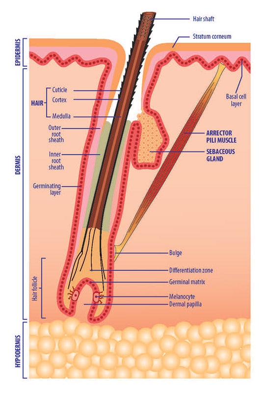
The mesenchymal compartment
It includes the dermal sheath and dermal papilla:
- The dermal sheath is an extracellular matrix composed mainly of type I and type III collagen and proteoglycans.
- The dermal papilla, a permanent unit at the base of the follicle, is composed of connective tissue and mesenchymal cells called dermal papillary fibroblasts. These cells specifically express alkaline phosphatase, CD133, the anti-apoptosis protein Bcl-2, cyclooxygenase type 1, PGE2 and CRABP1. The dermal papillary fibroblasts generate signals prompting proliferation, migration and differentiation of matrix cells, which are essential to the growth of the hair follicle [Assay: DPF-0002].
In addition to the cells of the follicular bulb, the fibroblasts of the dermal papilla could also act as a reservoir of pluripotent stem cells capable of differentiating into other cell lines: chondrocytes, osteoblasts, adipocytes, etc. The basal membrane ensures the separation of the epithelial and dermal compartments. It is rich in extracellular matrix, which is composed mainly of collagen IV, laminin V, fibronectin and heparin sulfate [Assay: FOLL-0003].
The sebaceous gland
The sebaceous gland is composed of sebocytes, which are epithelial cells forming the sebum. Sebum protects hair from dehydration and plays a role in the microbial ecosystem. Sebocytes express specific markers such as K7 and EMA and produce lipids (triglycerides, waxes, squalene, cholesterol), which they release by rupturing when differentiation is complete [Assays: SEBOAR-0004; SEBOAR-0007; SEBOAR-0019]. Deregulated sebum production can lead to dry or greasy hair. Apocrine sweat glands, which open into the top part of the hair follicles, are responsible for body odours associated with sweating.
Hair follicle: cell signalling and growth
There are four phases in the hair growth cycle. The transitions are carefully controlled by a complex set of soluble factor activators or inhibitors (growth factors, morphogens). These phases occur independently for each follicle, which ensures hair permanence.
The anagen phase is the phase during which the hair increases in length (by producing the hair shaft). This phase usually lasts for an average of 3 years, according to the individuals and to environmental factors. It applies to about 85% of hair follicles. The duration of this phase, which determines the length of the hair, depends on continued proliferation and differentiation of matrix cells [Assay: FOLL-0001 & FOLL-0002].
Hair growth kinetics [Assay: FOLL-0001]
The catagen phase is the transition between the anagen and telogen phases and is controlled by growth factors such as prostaglandin D2. This regression phase lasts an average of three weeks, during which hair growth stops. It progressively leads to a latency phase. During this phase, which applies to 1 to 2% of hair follicles , the epithelial cells at the base of the follicle enter apoptosis [Assay: FOLL-0002] while the cells of the dermal papilla remain intact and migrate close to the stem cells of the bulb to enter a latency phase.
The telogen phase, initially described as a latency phase that can last for up to three months, was redefined by using transcriptome studies. It applies to about 15 % of hair follicles.
The kenogen phase is a latency phase, which is not systematically observed in each cycle. It occurs after the hair falls out and lasts two to five months.
At the end of a cycle, all hair follicle compartments, with the exception of the dermal papilla, have been degraded by an apoptotic process. The start of a new cycle, recently named the neogen phase, is characterised by the regeneration of the follicle from a reservoir of pluripotent stem cells that proliferate and differentiate under the effects of various signalling pathways (BMP signalling pathway, Wnt/β-catenin signalling pathway). Other growth factors could block the initiation of this phase.


