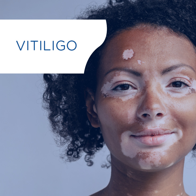
Skin pigmentation is much more than a cosmetic trait; it reflects profound biological differences that shape how the skin functions, ages, and responds to treatments. While the cosmetics industry has long relied on a one-size-fits-all approach, emerging evidence shows this model is inadequate. From variations in melanocyte activity to differential vulnerability to UV and visible light, understanding pigmentation diversity is essential for developing effective, safe, and culturally relevant skincare. This article explores why personalization is not just a trend but an imperative, and how science can help create solutions that truly respect the diversity of skin.
Discover our related blog articles
1. Understanding Skin Pigmentation Diversity
The biological mechanisms that determine skin color are far more intricate than they appear on the surface. From genetic variations to cellular interactions, pigmentation reflects a sophisticated interplay of factors that define both appearance and function.
1.1 A complex genetic architecture
Skin pigmentation is a highly polygenic trait shaped by thousands of years of evolution and adaptation to different environments. A recent cross-species curation has identified more than 650 genes associated with pigmentation phenotypes (https://www.ifpcs.org/colorgenes/). These genes contribute to melanocyte development, survival, pigment synthesis, and responses to environmental factors. In addition, large-scale gene expression studies have revealed hundreds of other genes that differ between skin types and likely influence pigmentation patterns and behaviors. This complex genetic landscape underlies the remarkable diversity of skin tones observed across populations worldwide.
1.2 Melanin types and distribution
At the cellular level, pigmentation is determined by the quantity, type, and distribution of melanin produced by melanocytes in the basal layer of the epidermis. Two main forms of melanin coexist in human skin: eumelanin, which confers brown to black coloration and provides effective photoprotection, and pheomelanin, which contributes yellow to red hues and generates more reactive oxygen species upon UV exposure. The proportion of these pigments varies widely between individuals and ethnic groups.
1.3 Cellular and microenvironmental interactions
Beyond melanocytes themselves, keratinocytes and fibroblasts also participate in regulating pigmentation. They influence melanogenesis through paracrine signals, oxidative stress modulation, and melanosome transfer to the upper layers of the epidermis. Research has highlighted significant differences in melanocyte morphology, dendricity, and enzyme activity depending on genetic background.
1.4 An evolutionary adaptation
Altogether, the structural and functional diversity of pigmentation reflects both inherited genetic factors and the adaptations that have enabled human skin to meet local environmental challenges. Recognizing this diversity is the first step toward developing personalized care solutions that respect the intrinsic complexity of differently pigmented skin.
2. Functional Implications of Pigmentation Diversity
Understanding the biology of skin pigmentation is not only a matter of genetics or cellular architecture, it also has tangible consequences for how skin behaves, how it interacts with its environment, and how it responds to topical products. These functional implications are essential for designing skincare solutions that are both effective and respectful of individual needs.
2.1. Differences in skin physiology
Diverse pigmentation phenotypes are associated with distinct physiological profiles that influence skin’s barrier function, hydration, and reactivity:
- Barrier integrity and transepidermal water loss (TEWL): The link between pigmentation and barrier function is complex. Some studies report higher TEWL in darker skin after disruption, while others find no difference or even lower TEWL. Notably, Reed et al. showed that lighter skin has lower stratum corneum integrity and slower barrier recovery. Liu et al. further demonstrated that in vitiligo lesions (devoid of melanin), barrier repair was significantly delayed despite similar baseline TEWL, suggesting that melanin and melanocyte signals actively support barrier homeostasis and recovery.
- Skin surface pH and barrier recovery dynamics: Pigmentation also influences the acid-base balance of the skin, which plays a critical role in enzymatic activity, microbiome stability, and overall cutaneous resilience. Baseline measurements show that the skin surface pH tends to be higher in individuals with darker skin, particularly among Black African populations compared to Caucasians. However, after barrier disruption, darker skin demonstrates a more pronounced decrease in pH, indicating faster reacidification. This accelerated acid mantle recovery may contribute to the superior barrier repair observed in melanin-rich skin. These pH dynamics further reinforce the notion that pigmentation modulates not only structural properties, but also functional recovery processes following external stress.
Beyond these biophysical parameters, pigmentation-related differences extend their influence to clinical manifestations, modulating how the skin reacts to inflammation, environmental stressors, or dermatological procedures.
2.2. Clinical consequences: Sensitivity, pigmentation disorders, and inflammatory responses
Pigmentation phenotype influences more than structural skin properties; it also shapes the clinical behavior of the skin, particularly in contexts of inflammation, environmental exposure, or cosmetic intervention:
- Post-inflammatory hyperpigmentation (PIH): Melanin-rich skin, particularly Fitzpatrick phototypes III to VI, is more prone to PIH following trauma, inflammation, or dermatological procedures. Even minimal irritation (such as exfoliation, acne lesions, or friction) can lead to persistent hyperpigmented marks, making product tolerance and gentle formulation especially important for these phototypes.
- Melasma susceptibility: Darker skin types also exhibit greater susceptibility to melasma, a chronic pigmentary condition characterized by symmetrical brown patches, especially on the face. While its exact pathogenesis is multifactorial, it is exacerbated by UV and visible light exposure, hormonal fluctuations, and inflammation, all factors that interact with melanin biology.
- Heightened reactivity in lighter skin types: Fair skin (phototypes I-II), with low eumelanin content, is less protected from UV and oxidative stress. This makes it more prone to acute erythema, stinging, and inflammatory flare-ups. Lighter phototypes show increased reactivity to environmental and topical aggressors and are more frequently affected by rosacea and contact dermatitis.
- Phototype-associated disorders: Certain skin conditions exhibit distinct prevalence and presentation depending on phototype. Acne and seborrheic dermatitis are more frequently observed in darker skin, where they tend to induce more visible and persistent post-inflammatory hyperpigmentation. In contrast, lighter phototypes are more commonly affected by vascular and inflammatory disorders such as rosacea and atopic dermatitis, often with greater erythema and sensory discomfort.
- Environmental burden and skin pigmentation: Emerging research also shows that individuals with darker skin are disproportionately exposed to urban air pollution, particularly fine particulate matter (PM2.5). This adds another dimension to pigmentary vulnerability: in addition to biological predispositions to hyperpigmentation, environmental inequalities increase the burden on melanin-rich skin, reinforcing the need for protective strategies adapted to these populations.
- Dermal senescence and pigmentary disorders: In melanin-rich skin, pigmentary conditions such as melasma are increasingly associated with alterations beyond the epidermis. Recent findings highlight a significant contribution of senescent dermal fibroblasts and mast cells to melanogenesis via pro-pigmentary cytokines and growth factors. This senescence-driven signalling may amplify or sustain hyperpigmentation even in the absence of acute inflammation or sun exposure. Addressing pigmentary disorders thus requires not only photoprotection and anti-inflammatory strategies, but also interventions targeting the dermal microenvironment and its aging status.
2.3. Aging: a phototype-dependent process
Skin aging is a complex interplay of intrinsic and extrinsic mechanisms that manifest differently depending on pigmentation type. Phototype influences not only the rate at which aging signs appear, but also their nature and distribution.
- In lighter skin (phototypes I–II), signs of aging typically emerge earlier and include prominent wrinkles, loss of firmness, and telangiectasia, especially in UV-exposed areas. This is largely due to the lower content of protective eumelanin, resulting in greater susceptibility to photoaging through cumulative oxidative stress and collagen degradation.
- In darker skin (phototypes IV–VI), chronological aging tends to be delayed, with better dermal density and collagen preservation. However, aging more often presents as dyschromia, uneven tone, or volume loss, sometimes accompanied by elastosis. Although wrinkles may be less marked, pigmentary changes are more frequent and impactful on perceived age.
Advanced imaging technologies such as those developed by QIMA Newtone have confirmed these distinct aging trajectories. In a comparative study, Campiche et al. demonstrated that wrinkle-related signs were more prominent in Caucasian and Asian skin, while African skin showed a greater burden of pigmentary heterogeneity and dark spots. These differences also reflect diverging consumer concerns: in lighter skin, aging is primarily associated with wrinkles, whereas in darker skin, tone evenness and brightness become the main focus. Such findings support the development of phototype-adapted assessment tools and reinforce the need for targeted anti-aging strategies that address both biological differences and cultural expectations.
2.4. Visible light and pigmentary aging: an underestimated threat
While UV radiation is well known for its impact on skin aging and pigmentation, emerging evidence highlights a critical role of visible light (especially blue light) in inducing hyperpigmentation, particularly in darker phototypes (IV to VI). Visible light promotes oxidative stress, melanin synthesis via opsin-3 signalling, and increases pigmentation even in the absence of UV exposure.
Tinted sunscreens containing mineral filters such as iron oxides remain the most effective option to protect against visible light. However, their formulation must be adapted to phototype preferences and optical needs. These findings reinforce the importance of phototype-specific photoprotection strategies — especially in the context of melasma or PIH management, where visible light plays a key role.
3. From Understanding to Action: The Case for Personalized Skincare
The diversity of skin physiology, clinical behavior, and aging trajectories across phototypes underscores a pressing need: skincare must evolve beyond standardized approaches to embrace biologically-informed, culturally-sensitive personalization.
3.1. Limitations of the one-size-fits-all model
Historically, most skincare products have been formulated and tested on a narrow range of skin types, often lighter phototypes of European descent. This bias has led to inappropriate product recommendations and underperformance, or even adverse effects, for individuals with different pigmentation profiles. For example, depigmenting agents or chemical exfoliants developed for fair skin may provoke post-inflammatory hyperpigmentation in darker skin types. Conversely, anti-aging formulas designed to reduce wrinkles may fail to address dyschromia, which represents a dominant concern in melanin-rich skin.
3.2. Toward evidence-based personalization
Personalized skincare does not imply anecdotal tailoring but must rely on scientifically validated differences. Recognizing the unique biophysical, biochemical, and perceptual features of each phototype enables a more targeted formulation strategy. This includes:
- Adapting active ingredients and concentrations to suit skin’s barrier profile, lipid composition, and sensitivity thresholds.
- Designing photoprotection tailored to phototype-specific vulnerabilities; for instance, including pigments to block visible light in darker skin, which is more susceptible to hyperpigmentation triggered by blue light.
- Prioritizing even tone and antioxidant support in darker skin, versus collagen stimulation and elasticity improvement in lighter phototypes.
3.3. Rethinking testing models and protocols
To truly serve diverse populations, personalization must begin in the laboratory. In vitro and ex vivo models should include keratinocytes and melanocytes from different phototypes to better predict responses to ingredients or formulations. Clinical studies must also recruit balanced panels representing a broad spectrum of skin colors, ethnicities, and environmental exposures.
At QIMA Life Sciences, this vision is already a reality. We support both finished product brands and active ingredient suppliers across all phases of evaluation, from preclinical modeling to clinical validation. Our dedicated in vitro and ex vivo platforms incorporate cells from multiple phototypes, enabling a more accurate prediction of efficacy and tolerance across diverse skin types. These models are particularly valuable for anticipating pigmentary risks such as post-inflammatory hyperpigmentation when testing exfoliants, depigmenting agents or photoprotective formulas. On the clinical side, we help design and conduct inclusive study protocols that are aligned with both the biological specificities and cultural expectations of global consumers.
Personalized skincare, in this sense, is not merely a market trend but a scientific and ethical imperative. It requires integrating dermatological biology, ethnodermatology, and consumer insight to deliver solutions that are not only effective, but also inclusive and respectful of individual needs.
4. Conclusion: Personalization as a Standard of Care
As scientific knowledge deepens, it becomes increasingly clear that skin pigmentation is not a superficial trait but a biologically meaningful parameter that shapes nearly every aspect of cutaneous function and response. The diversity of skin types across the globe demands an equally nuanced approach in skincare research, formulation, and clinical evaluation.
The transition from a generic, one-size-fits-all model to a personalized strategy is not only a question of product performance, but also a matter of dermatological relevance, consumer safety, and scientific integrity. Future skincare must acknowledge and integrate the biological realities of pigmentation, from cellular models to clinical endpoints, and from regulatory frameworks to marketing narratives.
Ultimately, building inclusive skincare means more than expanding shade ranges. It requires a paradigm shift that honors the complexity of human skin in all its diversity, bridging science and sensitivity, precision and perception. This evolution marks a pivotal step toward more respectful, equitable, and effective skincare solutions for all.
Written by:
Rachida Nachat-Kappes, PhD
Skin Biology Expert
Edited by:
Mara Carloni, PhD
Scientific Communications & Marketing Project Leader
Last update: 30/10/2025
Understanding how pigmentation shapes skin function and response is essential to designing inclusive, effective skincare. At QIMA Life Sciences, we offer advanced preclinical and clinical models that account for the diversity of phototypes and ethnicities.
With a comprehensive portfolio of in vitro, ex vivo and clinical platforms, we enable the evaluation of ingredient efficacy, safety, and tolerance across skin types representative of phototypes I to VI, helping you anticipate pigmentary risks and tailor your formulations accordingly.
From depigmenting agents to anti-(photo)aging or photoprotective solutions, we help brands move beyond one-size-fits-all skincare and embrace scientifically grounded personalization.
Eager to bring your skincare innovations to life?
References
- Baxter LL, Watkins‐Chow DE, Pavan WJ, and Loftus SK. A curated gene list for expanding the horizons of pigmentation biology. Pigment Cell Melanoma Res. 2019
- Naik PP and Farrukh SN. Influence of Ethnicities and Skin Color Variations in Different Populations: A Review. Skin Pharmacol Physiol. 2022
- Schlessinger DI, Anoruo M, and Schlessinger J. Biochemistry, Melanin. StatPearls. StatPearls Publishing: Treasure Island (FL); 2025.
- Upadhyay PR, Ho T, and Abdel-Malek ZA. Participation of keratinocyte- and fibroblast-derived factors in melanocyte homeostasis, the response to UV, and pigmentary disorders. Pigment Cell Melanoma Res. 2021
- Lucock MD. The evolution of human skin pigmentation: A changing medley of vitamins, genetic variability, and UV radiation during human expansion. Am J Biol Anthropol. 2023
- Wilson D, Berardesca E, and Maibach HI. In vitro transepidermal water loss: differences between black and white human skin. Br J Dermatol. 1988
- Kompaore F, Marty JP, and Dupont C. In vivo evaluation of the stratum corneum barrier function in blacks, Caucasians and Asians with two noninvasive methods. Skin Pharmacol. 1993
- Singh J, Gross M, Sage B, Davis HT, and Maibach HI. Effect of saline iontophoresis on skin barrier function and cutaneous irritation in four ethnic groups. Food Chem Toxicol. 2000
- Reed JT, Ghadially R, and Elias PM. Skin type, but neither race nor gender, influence epidermal permeability barrier function. Arch Dermatol. 1995
- Liu J, Man WY, Lv CZ, Song SP, Shi YJ, Elias PM, and Man MQ. Epidermal Permeability Barrier Recovery Is Delayed in Vitiligo-Involved Sites. Skin Pharmacol Physiol. 2010
- Fotoh C, Elkhyat A, Mac S, Sainthillier JM, and Humbert P. Cutaneous differences between Black, African or Caribbean Mixed-race and Caucasian women: biometrological approach of the hydrolipidic film. Skin Res Technol. 2008
- Berardesca E, Pirot F, Singh M, and Maibach H. Differences in stratum corneum pH gradient when comparing white Caucasian and black African-American skin. Br J Dermatol. 1998
- Lawrence E, Syed HA, and Al Aboud KM. Postinflammatory Hyperpigmentation. StatPearls. StatPearls Publishing: Treasure Island (FL); 2025.
- Mpofana N, Chibi B, Gqaleni N, Hussein A, Finlayson AJ, Kgarosi K, and Dlova NC. Melasma in people with darker skin types: a scoping review protocol on prevalence, treatment options for melasma and impact on quality of life. Syst Rev. 2023
- Passeron T, Lim HW, Goh C ‐L., Kang HY, Ly F, Morita A, Ocampo Candiani J, Puig S, Schalka S, Wei L, et al. Photoprotection according to skin phototype and dermatoses: practical recommendations from an expert panel. J Eur Acad Dermatol Venereol. 2021
- Tsuchida K, Sakiyama N, Ogura Y, and Kobayashi M. Skin lightness affects ultraviolet A-induced oxidative stress: Evaluation using ultraweak photon emission measurement. Exp Dermatol. 2023
- Litchman G, Nair PA, Atwater AR, and Bhutta BS. Contact Dermatitis. StatPearls. StatPearls Publishing: Treasure Island (FL); 2025.
- Alchorne MM de A, Conceição K da C, Barraza LL, and Milanez Morgado de Abreu MA. Dermatology in black skin. An Bras Dermatol. 2024
- Tan J and Berg M. Rosacea: current state of epidemiology. J Am Acad Dermatol. 2013;69(6 Suppl 1):S27-35.
- Aguilar-Gomez S, Cardenas JC, and Salas Diaz R. Environmental justice beyond race: Skin tone and exposure to air pollution. Proc Natl Acad Sci U S A. 2025
- Kim Y, Kang B, Kim JC, Park TJ, and Kang HY. Senescent Fibroblast-Derived GDF15 Induces Skin Pigmentation. J Invest Dermatol. 2020
- Venkatesh S, Maymone MBC, and Vashi NA. Aging in skin of color. Clinics in Dermatology. 2019
- Chien AL, Qi J, Grandhi R, Kim N, César SSA, Harris-Tryon T, Jang MS, Olowoyeye O, Kuhn D, Leung S, et al. Effect of Age, Gender, and Sun Exposure on Ethnic Skin Photoaging: Evidence Gathered Using a New Photonumeric Scale. J Natl Med Assoc. 2018
- Campiche R, Trevisan S, Séroul P, Rawlings AV, Adnet C, Imfeld D, and Voegeli R. Appearance of aging signs in differently pigmented facial skin by a novel imaging system. Journal of Cosmetic Dermatology. 2019
- Lim HW, Kohli I, Granger C, Trullàs C, Piquero-Casals J, Narda M, Masson P, Krutmann J, and Passeron T. Photoprotection of the Skin from Visible Light‒Induced Pigmentation: Current Testing Methods and Proposed Harmonization. J Invest Dermatol. 2021
- Setty SRG. Opsin3—A Link to Visible Light-Induced Skin Pigmentation. J Invest Dermatol. 2018


