

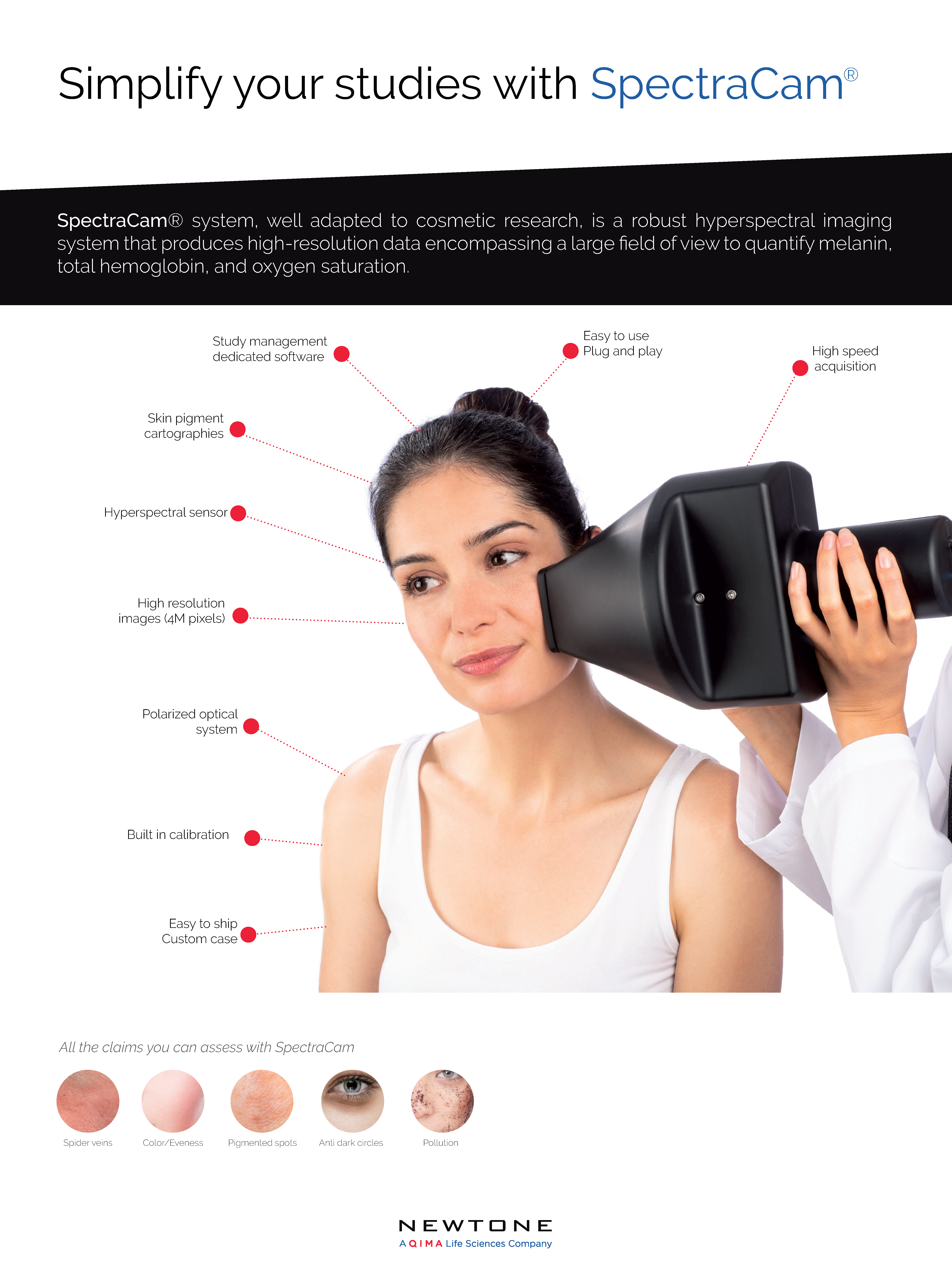
What is SpectraCam?
This is the first non-invasive polarized hyperspectral imaging system developed for cosmetic and dermatological studies. Unlike regular color images, SpectraCam allows for objective quantification of skin molecules located beneath the surface, such as total hemoglobin and melanin, and skin parameters, such as oxygen saturation.
Indeed, this portative system enables image acquisition all over the body revealing otherwise invisible parameters to the naked eye. As a result, you’ll get specific maps that visually represent the obtained results.
[Worth noting is that SpectraCam is suitable for use on phototypes I to IV.]
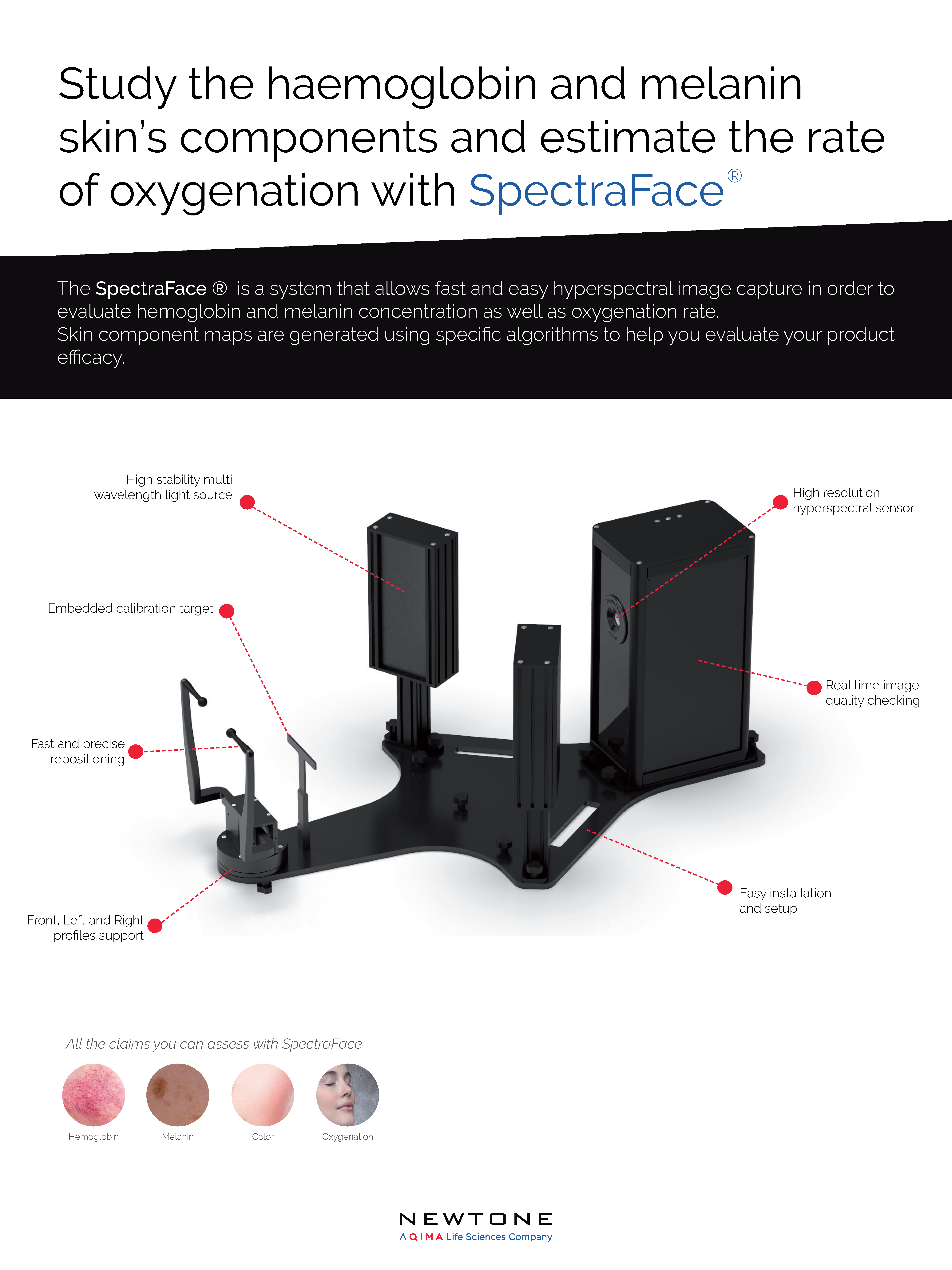
What is SpectraFace?
The SpectraFace system was designed to extend the capabilities of the SpectraCam from 2D to 3D imaging. While the SpectraCam excels in capturing images of flat skin areas, SpectraFace is optimized to address larger, non-flat areas, particularly the human face.
Functioning similarly to the SpectraCam, the SpectraFace system employs a non-invasive approach, capturing images at a social distance without physical contact. This unique device enables the generation of “mirror-like” full-face images, offering a comprehensive view of the facial skin components.
Download the SpectraCam
technical datasheet
How do they work?
Skin chromophores, like hemoglobin and melanin, are molecules that absorb particular wavelengths of visible light. As a result, they influence the perception of skin color and need to be taken into account when studying pigmentation conditions. These molecules are also relevant for the study of skin inflammation and vascularization.
The remarkable capabilities of SpectraCam and SpectraFace in quantifying these essential molecules and assessing skin pigmentation rely on their high-resolution cameras and the use of a hyperspectral system. Analyses of standard color images fail to precisely quantify chromophores and photophores located beneath the skin’s surface since they remain mostly invisible to the naked eye. The hyperspectral imaging approach overcomes this limitation by taking into account the specific spectral absorbance of these molecules.
Hyperspectral imaging involves capturing multiple images at various wavelengths across the visible spectrum, resulting in one spectrum per pixel in the image. By doing so, the concentrations of certain skin molecules, such as hemoglobin, melanin, and oxygen, can be estimated for each pixel. This valuable information is then presented in the form of images or maps, allowing for the observation of different structures like veins, blood capillaries, pores, hematomas, and pigmented spots.
Download the SpectraFace
technical datasheet
Why you should choose SpectraCam & SpectraFace
SKIN COMPONENT MAPS:
- Innovative algorithm for skin components qualification
- Visible reconstructed image generation
- Color analysis (L*, a*, b*, C, h)
- Melanin, hemoglobin, and oxygen saturation maps
- Skin component concentration on the entire acquired region or selected areas
- Precise detection of underlying structures
- Highly reproducible and validated process
INNOVATIVE HYPERSPECTRAL ACQUISITION:
- Full visible spectrum: 400 to 700 nm with 10 nm bandwidth
- Innovative lightening system with multiband LEDs
- Polarized optical module to reduce specular reflection
- Build-in calibration with specific black and white samples
- Simple: configured and guided system
- Quick: capture time with full spectrum in less than 5 seconds
- Precise: high-resolution sensor
- Connected: automatic image transfer to the Newtone cloud
Investigate skin components beneath the skin surface & substantiate your claims

Skin parameters
• Skin thickness
• Melanin volume fraction
• Blood volume fraction
• Oxygen saturation
Associated claims
• Skin color and eveness
• Skin pigmentation
• Pigmented spots
• Anti-dark circles
• Hematomas
• Skin oxygenation
• Skin inflammation
• Skin redness
• Spider veins
• Pollution
You may also be interested in
ColorFace®

Related Publications & Articles
Gevaux L., Gierschendorf J., Rangot J., Cherel M., Séroul P., Nkengne A., Robic J., Trémeau A., Hébert M. Real-time skin chromophore estimation from hyperspectral images using a neural network. Skin Research and Technology (2020).
Vergnaud, H., Cherel, M., François, G., Charton, Z., Loescher, E., Caisey, L., Gazano, G. Lip color measurement: A new hyperspectral imaging device. Skin Research and Technology (2023).
More resources here
