in vitro models & assays
QIMA Life Sciences has many in vitro models and ex vivo models at your disposal:
- intrinsic ageing model:
- “aged” human dermal fibroblasts (Hayflick model)
- “aged” dermal equivalent (Hayflick model)
- ex vivo human skin aging models
- accelerated ageing model:
- human dermal fibroblasts aged by oxidative stress (H2O2)
- human reconstructed skin in deficient medium
- photo-ageing model:
- human dermal fibroblasts subjected to UVA, infrared or UV radiation
- photo-aged human full thickness reconstructed skin
on which we can evaluate the anti-ageing effect of active cosmetic compounds on:
- cell renewal (cell proliferation, migration and differentiation)
- extracellular matrix synthesis and degradation (collagen, elastin, hyaluronic acid, MMPs, etc.)
- senescence marker expression
- DNA damage
- free radical production
- mitochondrial biogenesis & energy metabolism
- Expression and activities of anti-oxidant enzymes
- Expression and activation of anti-oxidant transcription factors and proteins
- Lipid peroxidation
- Protein carbonylation
Here are a few examples among all standard assays proposed by QIMA Life Sciences in the field of skin ageing:
Biochemical analysis of non-invasive clinical samples
Analysis of lipids involved in the barrier function
Our company has developed ready-to-use non-invasive collection kits to analyze the lipids and biomarkers of the skin surface from your samples or from those of your clinical center.
The epidermal lipids involved in the barrier function of the epidermis (ceramides, fatty acids and cholesterol) are removed using the SW Kit.
The analysis of these lipids makes it possible to evaluate the quality of the intercorneocyte cement involved in the barrier function of the epidermis and in the prevention of transepidermal water loss (TEWL).
These evaluations help support your claims about the efficacy of biomimetic products, barrier products, protective products, moisturizers, etc.
Analysis of the components of the Natural Moisturizing Factor (NMF)
The amino acids and minerals present on the surface of the skin are collected using the SW Kit:
- PCA / UCA (cis/trans) – (catabolites of filaggrin)
- Amino acids
- Urea, lactates
- Mineral elements: Ca, K, Na, Mg, Zn, etc.
The analysis of these compounds makes it possible to evaluate the impact of the NMF component on skin hydration
These analysis help support your claims about the efficacy of hygroscopic products, barrier products, protective products, moisturizers, etc.
Analysis of markers of oxidative stress
Oxidative stress is also a factor of accelerated skin ageing. Oxidative stress markers are analyzed from samples using the SW Kit. The sampling areas depend on the type of stress (induced stress or external environment).
The analyzed markers are:
- products from peroxidation and lipid detoxification (MDA, peroxidized squalene, CAT, SOD, etc.)
- protein oxidation products (AOPP Dityrosin, ROH, etc.)
These evaluations help support your claims about the efficacy of anti-pollution products, protective products , anti-aging products, etc.

Screening des céramides – LC/MS
–
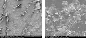
Cornéocytes endommagés et sains – SEMX500
–
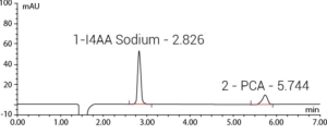
Analyse des PCA – LC/UV
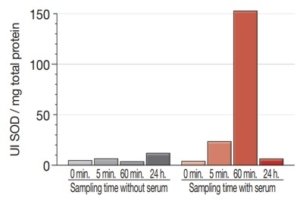
Quantification de la SOD
Data mapping and clinical imaging
Measurement of wrinkles, fine lines, and skin roughness, on whole face, in 2D imaging
On the high-resolution image of the whole face, image analysis makes it possible to monitor severity markers of visible wrinkles and fine lines.
Length, surface, depth, and projected volume are parameters that can be extracted to monitor the relief evolution induced by a treatment.
Image analysis algorithms are adapted to the required threshold of detection (first or marked wrinkles, fine lines or very small, oriented elements of the microrelief) and so can be used for any type of topic (wrinkles of a given area or of the whole face, different types of ethnicities with different types of wrinkles).
Newtone Technologies has developed specific algorithms for eye contour, lips, glabella, as well as for cheeks and nasolabial folds. These algorithms can be adapted to Caucasian, Asian, Hispanic, and African panels.
Roughness parameters can be extracted by image analysis and the evolution of skin smoothness can be monitored. 3D images can also be analyzed and can provide integrated parameters of roughness for the investigated surface.
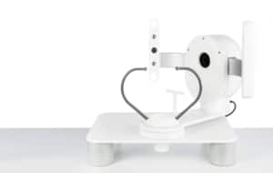
ColorFace®
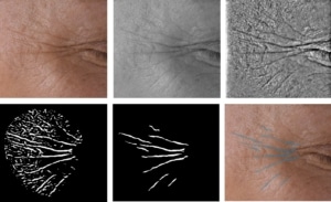 Process of analysis of eye contour wrinkles
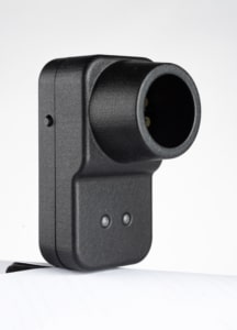
Newtone SkinCam
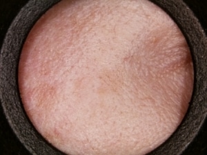
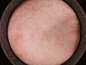
Image acquisition of temple with SkinCam®
Measurement of pigment signs of sun ageing (lentigo) in 2 D
Skin ageing signs, such as solar lentigos, can be detected and analyzed by image analysis, on the whole face, on the back of the hand or by targeting a cutaneous lesion.
According to the area to be investigated, a choice will be made regarding the type of shot for the whole face, the hand, or a small sun-exposed area of the body (e.g., the upper chest).
The density of pigmentation spots, their surface, their color intensity, and the contrast measured with surrounding skin, as well as homogeneity of complexion are all parameters that can be extracted from this analysis.
ColorFace®
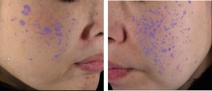
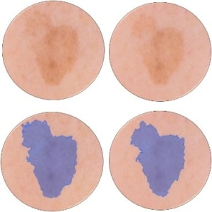
Analysis of solar lentigos on the whole face, with images acquired with ColorFace®

DigiCam

Image acquisition of back of hand with DigiCam® and analysis of pigmentation spots

SkinCam
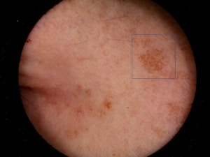 Image acquisition of temple with SkinCam®
Measurement of non-visible pigmentation in UV imaging
Investigation of sun-induced pigmentation, which is non-invisible but will appear with age, can be performed by using UV polarized monochrome imaging. Parameters such as lesion surface and density can be extracted and monitored over time.
Measurement of melanin in hyperspectral imaging
Newtone’s hyperspectral imaging is a unique technology developed to calculate the skin chromophore absorption mappings, and consequently to measure evolution over time, while providing a perfectly representative image of the measured evolution.
Acquisition systems allow image acquisition of the whole face (SpectraFace) or of a defined area of the body and face (SpectraCam).
On these images, pigment lesions can be monitored and analyzed as by color imaging, while discriminating melanin and hemoglobin.
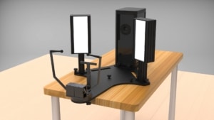
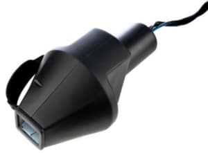
Hyperspectral imaging






in vitro modeling of skin photoaging: development of evaluation tools for cosmetics
Cosmetics_cat_publi, Skin ageingDevelopment of in vitro models skin for better understand the modifications during photo-aging induced by repetitive UV exposure.
Development of a new model of reconstructed aged skin useful to study antiageing effects of cosmetic compounds
Cell and tissue engineering, Skin ageingThe development of new anti-ageing products needs performant in vitro models mimicking morphological changes and physiological modifications appearing during skin ageing. In order to have access to a simple model mimicking the epidermis ageing but in relation with a normal dermis, we have developed a new in vitro model of reconstructed skin comprising an aged epidermis covering a reconstructed dermis built with collagen and normal (young) fibroblasts.
Effect of C-xyloside on morphogenesis of the dermal epidermal junction in aged female skin. An ultrastuctural pilot study
Cosmetics_cat_publi, Cosmétique, Skin ageingThese data suggest that topical C-xyloside application in vivo may be efficient in inducing a better dermal-epidermal cohesion when such a junction is deficient, as is the case in photo-aged or chronologically aged skin. Moreover, a statistically significant increase in CD44 expression was noted in the epidermis of C-xyloside-treated compared to the placebo treated skin areas.
Reduced expression of the adhesion protein tensin1 in cultured human dermal fibroblasts affects collagen gel contraction
Cosmetics_cat_publi, Cosmétique, Skin ageingtensin1 plays an important role in cell-matrix interaction and that a reduced expression might contribute to the dermal alterations observed during skin ageing