
 https://qima-lifesciences.com/wp-content/uploads/2019/09/190905_ESDR2019_Fig2.jpg
436
995
Vanessa
https://qima-lifesciences.com/wp-content/uploads/2023/03/QIMA_life_sciences.png
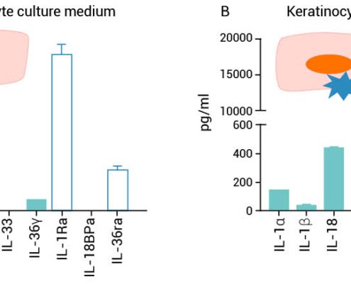
Vanessa2019-10-01 14:11:432019-10-07 08:27:51Fibroblasts are potentially the key sensors of epidermis lesion through keratinocyte IL-1 signalling
https://qima-lifesciences.com/wp-content/uploads/2019/09/190905_ESDR2019_Fig2.jpg
436
995
Vanessa
https://qima-lifesciences.com/wp-content/uploads/2023/03/QIMA_life_sciences.png
Vanessa2019-10-01 14:11:432019-10-07 08:27:51Fibroblasts are potentially the key sensors of epidermis lesion through keratinocyte IL-1 signalling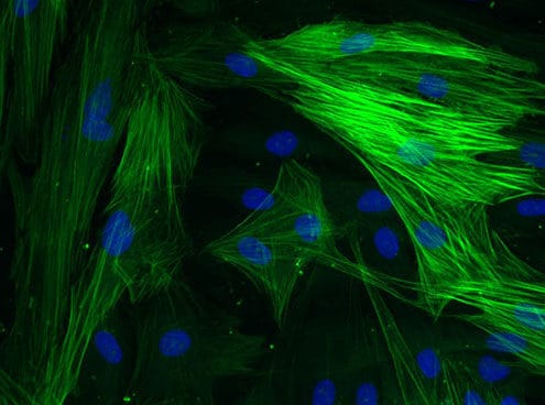 https://qima-lifesciences.com/wp-content/uploads/2016/11/mw_SMA.jpg
368
655
Admin istrateur
https://qima-lifesciences.com/wp-content/uploads/2023/03/QIMA_life_sciences.png
Admin istrateur2016-11-12 15:36:082025-03-20 14:24:04Cicatrisation cutanée, phases de granulation et maturation
https://qima-lifesciences.com/wp-content/uploads/2016/11/mw_SMA.jpg
368
655
Admin istrateur
https://qima-lifesciences.com/wp-content/uploads/2023/03/QIMA_life_sciences.png
Admin istrateur2016-11-12 15:36:082025-03-20 14:24:04Cicatrisation cutanée, phases de granulation et maturation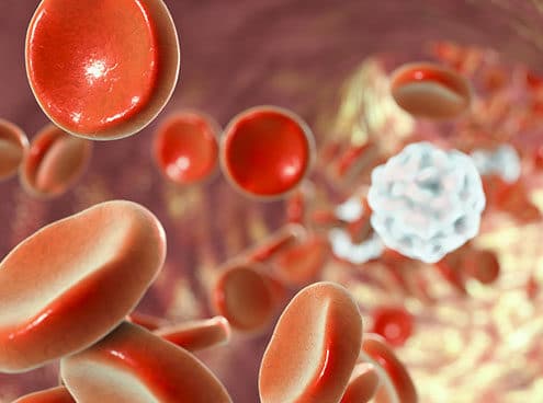 https://qima-lifesciences.com/wp-content/uploads/2016/11/mw_blood-cells-cicatrisation-inflammation-AdobeStock_117910248.jpg
368
655
Admin istrateur
https://qima-lifesciences.com/wp-content/uploads/2023/03/QIMA_life_sciences.png
Admin istrateur2016-11-10 15:40:392025-03-20 14:25:49Cicatrisation cutanée, hémostase et phase inflammatoire
https://qima-lifesciences.com/wp-content/uploads/2016/11/mw_blood-cells-cicatrisation-inflammation-AdobeStock_117910248.jpg
368
655
Admin istrateur
https://qima-lifesciences.com/wp-content/uploads/2023/03/QIMA_life_sciences.png
Admin istrateur2016-11-10 15:40:392025-03-20 14:25:49Cicatrisation cutanée, hémostase et phase inflammatoire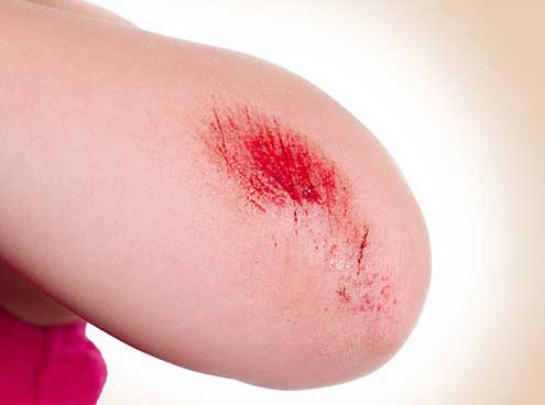 https://qima-lifesciences.com/wp-content/uploads/2016/03/pf_Cicatrisation-Adobe.jpg
368
655
Admin istrateur
https://qima-lifesciences.com/wp-content/uploads/2023/03/QIMA_life_sciences.png
Admin istrateur2016-11-09 15:45:552025-03-20 14:26:36Cicatrisation cutanée, généralités
https://qima-lifesciences.com/wp-content/uploads/2016/03/pf_Cicatrisation-Adobe.jpg
368
655
Admin istrateur
https://qima-lifesciences.com/wp-content/uploads/2023/03/QIMA_life_sciences.png
Admin istrateur2016-11-09 15:45:552025-03-20 14:26:36Cicatrisation cutanée, généralités https://qima-lifesciences.com/wp-content/uploads/2016/03/mw_study-of-proliferation-and-3D-epidermal-reconstruction-from-foreskin.jpg
368
655
Admin istrateur
https://qima-lifesciences.com/wp-content/uploads/2023/03/QIMA_life_sciences.png
Admin istrateur2014-09-16 16:18:492018-10-24 21:59:38Study of proliferation and 3D epidermal reconstruction from foreskin, auricular and trunk keratinocytes in children
https://qima-lifesciences.com/wp-content/uploads/2016/03/mw_study-of-proliferation-and-3D-epidermal-reconstruction-from-foreskin.jpg
368
655
Admin istrateur
https://qima-lifesciences.com/wp-content/uploads/2023/03/QIMA_life_sciences.png
Admin istrateur2014-09-16 16:18:492018-10-24 21:59:38Study of proliferation and 3D epidermal reconstruction from foreskin, auricular and trunk keratinocytes in children https://qima-lifesciences.com/wp-content/uploads/2016/03/mw_epidermal-healing-in-burns.jpg
368
655
Admin istrateur
https://qima-lifesciences.com/wp-content/uploads/2023/03/QIMA_life_sciences.png
Admin istrateur2014-09-07 16:13:052018-10-24 21:59:50Epidermal healing in burns: autologous keratinocyte transplantation as a standard procedure: update and perspective
https://qima-lifesciences.com/wp-content/uploads/2016/03/mw_epidermal-healing-in-burns.jpg
368
655
Admin istrateur
https://qima-lifesciences.com/wp-content/uploads/2023/03/QIMA_life_sciences.png
Admin istrateur2014-09-07 16:13:052018-10-24 21:59:50Epidermal healing in burns: autologous keratinocyte transplantation as a standard procedure: update and perspective https://qima-lifesciences.com/wp-content/uploads/2016/03/mw_foreskin-isolated-keratinocytes-provide.jpg
368
655
Admin istrateur
https://qima-lifesciences.com/wp-content/uploads/2023/03/QIMA_life_sciences.png
Admin istrateur2013-03-14 16:32:272018-10-24 22:01:55Foreskin-isolated keratinocytes provide successful extemporaneous autologous paediatric skin grafts
https://qima-lifesciences.com/wp-content/uploads/2016/03/mw_foreskin-isolated-keratinocytes-provide.jpg
368
655
Admin istrateur
https://qima-lifesciences.com/wp-content/uploads/2023/03/QIMA_life_sciences.png
Admin istrateur2013-03-14 16:32:272018-10-24 22:01:55Foreskin-isolated keratinocytes provide successful extemporaneous autologous paediatric skin grafts https://qima-lifesciences.com/wp-content/uploads/2016/05/mw_Foreskin-in-children.jpg
368
655
Admin istrateur
https://qima-lifesciences.com/wp-content/uploads/2023/03/QIMA_life_sciences.png
Admin istrateur2010-12-10 15:11:492018-10-24 22:04:20Quantitative and qualitative study in keratinocytes from foreskin in children: Perspective application in paediatric burns
https://qima-lifesciences.com/wp-content/uploads/2016/05/mw_Foreskin-in-children.jpg
368
655
Admin istrateur
https://qima-lifesciences.com/wp-content/uploads/2023/03/QIMA_life_sciences.png
Admin istrateur2010-12-10 15:11:492018-10-24 22:04:20Quantitative and qualitative study in keratinocytes from foreskin in children: Perspective application in paediatric burns https://qima-lifesciences.com/wp-content/uploads/2016/05/mw_Cultured-keratinocyte-cells-from-foreskin.jpg
368
655
Admin istrateur
https://qima-lifesciences.com/wp-content/uploads/2023/03/QIMA_life_sciences.png
Admin istrateur2009-02-04 13:56:522018-10-24 22:05:32Cultured keratinocyte cells from foreskin and future application for burns in children
https://qima-lifesciences.com/wp-content/uploads/2016/05/mw_Cultured-keratinocyte-cells-from-foreskin.jpg
368
655
Admin istrateur
https://qima-lifesciences.com/wp-content/uploads/2023/03/QIMA_life_sciences.png
Admin istrateur2009-02-04 13:56:522018-10-24 22:05:32Cultured keratinocyte cells from foreskin and future application for burns in children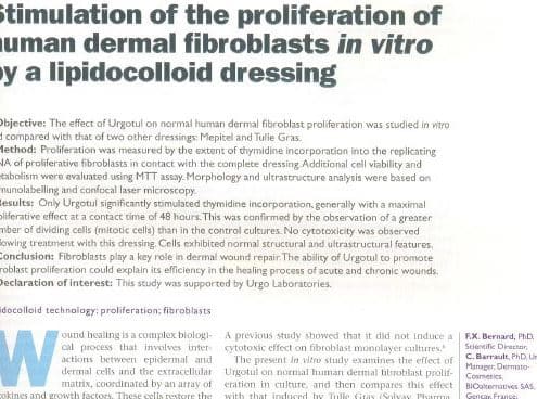 https://qima-lifesciences.com/wp-content/uploads/2016/05/mw_Lipidocolloid-dressing.jpg
368
655
Admin istrateur
https://qima-lifesciences.com/wp-content/uploads/2023/03/QIMA_life_sciences.png
Admin istrateur2005-05-15 10:59:162018-10-24 22:14:08Stimulation of the proliferation of human dermal fibroblasts in vitro by a lipidocolloid dressing
https://qima-lifesciences.com/wp-content/uploads/2016/05/mw_Lipidocolloid-dressing.jpg
368
655
Admin istrateur
https://qima-lifesciences.com/wp-content/uploads/2023/03/QIMA_life_sciences.png
Admin istrateur2005-05-15 10:59:162018-10-24 22:14:08Stimulation of the proliferation of human dermal fibroblasts in vitro by a lipidocolloid dressing